Investigations into the workings of the human brain are being undertaken by scientists from many disciplines. The Cardiff University Brain Research Imaging Centre (CUBRIC) aims to bring researchers together, offering a focus for physicists, bioengineers, neuroscientists, psychologists, psychiatrists, and computer scientists to access state-of-the-art brain imaging instruments.
CUBRIC was established in 2006 as part of Cardiff University's School of Psychology. Its remit is to investigate normal brain function and, increasingly, to study how this function is disrupted by the onset of diseases such as epilepsy, Alzheimer's, and schizophrenia. Medicalphysicsweb editor Tami Freeman visited the center to find out more.
MEG and EEG
Researchers at the Cardiff University Brain Research Imaging Centre can exploit a range of imaging techniques to study the workings of the human brain, using a wide array of imaging modalities.
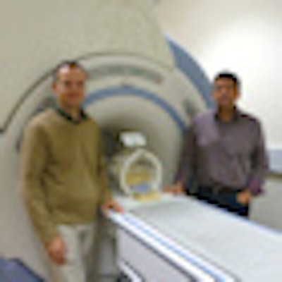
 Richard Wise and Krish Singh by CUBRIC's mock MRI system. Equipped with stimulus delivery systems, this system is used to test experimental ideas on volunteers in a realistic imaging environment, as well as helping them to acclimatize to the MRI environment.
One of CUBRIC's key research tools is magnetoencephalography (MEG), which measures the magnetic fields generated by neurons firing in the brain during specific cognitive tasks. These fields are detected using an array of pick-up coils located within a helmet surrounding the subject's head. As neural magnetic fields are extremely weak, of the order of 10-12 T, MEG systems employ extremely sensitive magnetic sensors called superconducting quantum-interference devices (SQUIDs).
CUBRIC's MEG system contains 275 SQUID channels that simultaneously measure the magnetic field around the entire head. The system resides in a shielded room to prevent interference from external fields (such as those induced by cars driving past), while additional SQUIDS measure the background environmental field for subtraction from the recorded brain signals.
"The good thing about MEG is that it measures the direct electrical activity of brain with very high temporal resolution," explained CUBRIC's MEG Director Krish Singh, PhD. "The disadvantage is that as the measurements are external, it requires solving of a difficult inverse problem to work out where in the brain the activity was located."
MEG is being used at CUBRIC to investigate areas including childhood epilepsy, Alzheimer's, memory processes, schizophrenia, and visual processing. When medicalphysicsweb visited, the researchers were performing a memory experiment in which a volunteer viewed a series of words displayed on a screen within the MEG system. While the subject recalled information regarding each word, indicating her responses using fiber-optic button boxes, the MEG system continuously recorded 275 traces from her brain.
Following the experiment, the signals recorded in the two seconds after each word is displayed will be analyzed and correlated with the accuracy of the subject's responses. "Data analysis is the big challenge," Singh said. He added that it's important to be able to break down the data into frequency ranges and perform a time-frequency analysis, another challenging task. As a result, the center is about to upgrade its processing power by installing a 1296 core computer cluster.
CUBRIC also has several dedicated labs for electroencephalography (EEG), a more established technique that uses electrodes attached to the scalp to measure voltage differences generated by the underlying neural activity. The researchers are using EEG mainly to identify electrical processes associated with different stages of memory function. EEG offers a similar temporal resolution to MEG, in the order of milliseconds, but lower spatial resolution.
MR and more
For MR-based studies of the brain, CUBRIC is equipped with a state-of-the-art 3-tesla MRI system located in a Faraday cage, both to isolate the system from outside interference and to confine the emitted fields within the room. The system is used for cognitive neuroscience and clinical research projects, employing both structural and functional MRI (fMRI).
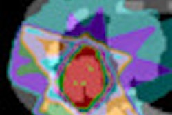
Structural scans using diffusion tension imaging (DTI), for example, can reveal connectivity between one part of the brain and another. DTI works by tracking the motion of water molecules, which move preferentially along white-matter tracts. "We are using diffusion imaging to look at the paths of fibers tracts in the brain, to determine how that wiring changes with diseases, as well as studying normal variability," explained Richard Wise, PhD, CUBRIC's fMRI Director.
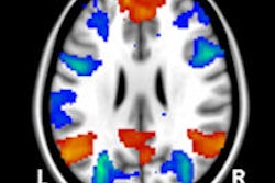
The other major tool is fMRI, which relies upon the measurement of blood oxygen levels. When the brain performs a task, areas associated with that task exhibit local increases in blood flow and oxygenation, which are detected as an increased MR signal. For functional studies, the CUBRIC MRI system is equipped with an integral video screen to display stimuli, as well as button boxes that enable subjects to indicate responses.
Functional MRI offers submillimeter spatial resolution and can record an entire brain scan every three seconds. This three-second temporal resolution is, however, lower than that offered by MEG and EGG, as the MR signal is derived from changes in blood flow, which are slower than neural activity.
To combine the advantages of both modalities, the CUBRIC team can perform EEG and MRI simultaneously, with the subject wearing an EEG cap within the MR system. This setup obviously introduces several challenges. For example, the MR field could induce currents in the EEG electrodes, leading to heating. This can be avoided via the use of resistors to avoid excessive currents.
Another potential complication is that the system's varying magnetic field induces large voltage artifacts in the EEG system. "You can end up with 10 mV artifacts, while trying to detect microvolts from the brain," Wise said. To address this, the researchers exploit the fact that scanner interference is repeatable, and subtract an average artifact from the detected signal. "It's a solved problem," Wise told medicalphysicsweb.
CUBRIC researchers recently used this simultaneous EEG/MRI approach to study the impact of caffeine on the brain. A 32-channel EEG system measured scalp potentials to determine neural effects, while fMRI revealed vascular changes. Another project involved the study of sedation, with EEG showing electrical activation and MRI revealing the brain blood flow.
Stimulation studies
The above techniques can all be thought of as correlative: A task is performed, changes in brain signal are monitored, and attempts are made to relate the two. The other experimental technique employed at CUBRIC -- transcranial magnetic stimulation (TMS) -- is slightly different. Here, a rapidly changing magnetic field, generated by electrical coils outside the head, is applied to induce weak electric currents in the brain.
The idea is to see how cognitive performance is affected when the coil is moved to different parts of the head, stimulating different areas of brain tissue. "Besides giving drugs, TMS is the only technique that we have for interfering with brain activity," Wise explained. "So it's extremely useful as a probe of what is going on."
The researchers are currently studying how TMS disrupts network function in the brain, to help elucidate how different areas of the brain communicate with each other. Here again they have developed a method for performing TMS within the MR system, using MRI to track the position of the coil with respect to various structures, in order to target specific regions of the brain.
Such an arrangement generates enormous forces on the TMS coil, and there are many challenges in getting it to work without breaking the coil, Wise explained. He noted that CUBRIC encourages its researchers to apply more than one modality to answer specific research questions, and to balance out strengths and weaknesses using a multimodal approach.
Currently, around 10 academics, 40 doctoral students, and 25 postdoctoral researchers regularly use the facilities at CUBRIC. According to Singh, the center is already running at full capacity and, five years after instigation, is now looking to expand.
This year, CUBRIC added a Master of Science degree in neuroimaging methods and applications to its academic degree programs. This is targeted to individuals who have a psychology/neuroscience background and physicists, mathematicians, or engineers interested in developing the next generation of methods.
© IOP Publishing Limited. Republished with permission from medicalphysicsweb, a community website covering fundamental research and emerging technologies in medical imaging and radiation therapy.