Multiscale imaging of the angiogenic network is uncovering new insights into tumor biology and challenging accepted anatomical models, but better tools -- both qualitative and validated -- are vital for multiparametric high-resolution mapping of structural, functional, cellular, and molecular imaging.
That's the view of Dr. Michal Neeman, PhD, dean of biology and director of the Clore Center for Biological Physics, Weizmann Institute of Science, Rehovot, Israel, who was a keynote speaker on Saturday at the World Molecular Imaging Congress (WMIC).
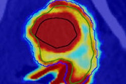
 New tools are necessary for multiparametric high-resolution mapping of structural, functional, cellular, and molecular imaging, according to Dr. Michal Neeman, PhD. All images courtesy of Brendan Lyon, ImageBureau, imagebureau.ie.
Environmental cues that set the stage for vasculogenesis and angiogenesis are complex and still not fully uncovered, she noted. Included here are hypoxia, extracellular matrix, oxidative stress, glucose, and hormones.
"Vascular maturation is critical and requires accessory cells," Neeman said. "Hemodynamic responses in a complicated vascular network can yield complex phenotypes. Vascular function is important."
She described recent work using arterial spin labeling MRI to enable the unique identification of individual fetuses in a pregnant mouse. This technique, which was described in the August 2012 issue of Magnetic Resonance in Medicine, allows for noninvasive, longitudinal studies of pregnant animals. A recent -- and as yet unpublished -- observation also suggests blood flow into individual placentas does not fit with the published anatomical model, she added.
"We need to redraw the anatomy of maternal vasculature in pregnant mice," she told WMIC attendees.
Neeman's group has developed several other novel MRI methods for imaging the key events during angiogenesis -- or the growth of new blood vessels -- during pregnancy and during the development of ovarian carcinomas.
The development of ovarian cancer is associated with the hormonal changes that accompany menopause, even though events during ovulation give rise to the initial risk. A thin layer of outer epithelial cells undergoes constant rupture to release oocytes for possible fertilization. In order to repair the repeated 'wound,' these epithelial cells do not undergo senescence, but are able to proliferate indefinitely, which gives rise to a cancer risk. Induction of an angiogenic switch is one of the key events driving tumor development.
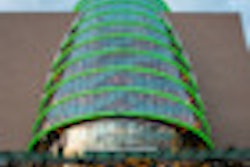
 Neeman and her colleagues are using arterial spin labeling MRI to enable the unique identification of individual fetuses in a pregnant mouse.
"Angiogenesis starts quite rapidly," Neeman commented. "You get this initial sprouting of angiogenesis."
Stabilization of the immature vascular network is a slower and more complicated process, but it is necessary for the tumor to progress. Part of that stabilization process involves the recruitment of infiltrating fibroblasts, which aid angiogenesis by depositing collagen and imparting structural solidity to the developing tumor vasculature.
Neeman and colleagues have developed an MRI-based technique for imaging in vivo gene expression using the intracellular iron storage protein ferritin as a reporter gene. This dispenses with the need to supply an exogenous contrast agent, while offering MRI-level sensitivity. Her group has used this approach to obtain quantitative data -- which is not yet published -- on fibroblast infiltration of ovarian tumors. Measuring this phenomenon could offer a means of assessing the efficacy of candidate therapies that target angiogenesis.
The Israeli team has deployed other imaging techniques for analyzing the key steps in angiogenesis and vascular functioning, including noninvasive macromolecular dynamic contrast-enhanced MRI, blood-oxygen-level-dependent (BOLD) MRI, and intravital two photon microscopy. Additional advances in imaging are required to further unpick the processes involved, Neeman concluded.