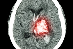
CT radiomics enables clinicians to predict the histological type and grade of retroperitoneal sarcomas more accurately, a U.K.-led study published in the November issue of The Lancet Oncology has found.
The results could have "important implications for improving diagnosis and risk stratification" of these cancers, wrote first author Dr. Amani Arthur, registrar at the Royal Marsden NHS Foundation Trust and clinical research fellow at the Institute of Cancer Research in London.
"Our radiomics model to predict histological subtype and grade presents a conceptual advance and an opportunity for a change in personalized care for patients with retroperitoneal sarcoma," the authors noted.
Retroperitoneal sarcomas are large and complex. They make up 12% to 15% of all soft tissue sarcomas, and patients with these sarcomas tend to have poor prognoses, the researchers explained. That's why the "clinical tools available for the upfront determination of histological type and grade urgently need to be improved to allow for improved risk stratification and management," they wrote -- and why CT radiomics could help.
Arthur and her colleagues sought to develop and validate a CT-based radiomics classification model for predicting histological type and grade of retroperitoneal leiomyosarcoma and liposarcoma. The team conducted a study they call Radiomics in Sarcoma of the Retroperitoneum (RADSARC-R), using "discovery" data taken from 170 patients at the Royal Marsden Hospital and validation data from 89 patients who participated in the phase III STRASS study of neoadjuvant radiotherapy in retroperitoneal sarcoma.
The STRASS study was conducted at a number of European centers as well as the Dana-Farber Cancer Institute in Boston. Study participants were adults with confirmed primary leiomyosarcoma or liposarcoma proceeding to surgical resection who had available contrast-enhanced CT scans (The Lancet Oncology, November 2023, Vol. 24: 11, pp. 1277-1286).
The researchers used the "discovery" information to create a CT-based radiomics workflow that included manual delineation, sub-segmentation, feature extraction, and predictive model building; they also built and tested classifiers to help predict histological type and low- versus intermediate- or high-grade tumor types.
The investigators found the following:
- The highest-performing model on validation for predicting the two most common histological types (liposarcoma and leiomyosarcoma) had an area under the receiver operator curve (AUROC) of 0.93; this model was based on a feature set of radiomics and radiomic volume fractions.
- The highest-performing model on validation for predicting histological grade had an AUROC of 0.88; this model was based on a radiomics feature set.
The findings could have "important implications for improving diagnosis and risk stratification in retroperitoneal sarcomas," the authors noted.
"These models could be further developed to address the intricate complexities of intermediate and higher-grade retroperitoneal sarcomas and for exploring the value of combining other radiological features or clinical data to the predictive performance of the models," they concluded.
The complete study can be found here.