MRI is the most accurate imaging modality for detecting a breast implant rupture, while mammography is the least sensitive yet the most specific method, and ultrasound is often used as a first-line screening tool but sensitivity is limited, according to award-winning research from a top London teaching hospital.
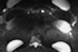
Prominent folds and reverberation artifacts on ultrasound can mislead the operator into suspecting an intracapsular rupture; silicone on either side of a continuous curvilinear hypointense structure within the implant on MRI is simply due to a prominent fold and not rupture, noted Dr. Philip Dilks, a radiologist from St. Bartholomew's Hospital. Meanwhile, fluid between the implant shell and capsule may be due to infection; correlation with clinical history and careful evaluation of the implant shell and characteristics of the surrounding fluid can help distinguish between infection and intracapsular rupture.
"The 'keyhole' sign is subtle and important to recognize as this may be the only sign of gel bleed or intracapsular rupture," he pointed out in an e-poster that received a certificate of merit at RSNA 2012. "Capsular contracture usually occurs within the first few months after surgery and is caused by contraction of the fibrous capsule. This results in a change in the shape of the implant and is usually a clinical diagnosis."
Small foci of water signal within a single lumen silicone implant is a nonspecific finding and does not necessarily indicate rupture, but this finding in a double lumen implant suggests rupture of the inner lumen and mixing of saline and silicone. Hyperintense T1 and T2 signal adjacent to membrane fragments within an implant can occur as a result of a chemical shift artifact and should not be confused with water type signal, Dilks continued.
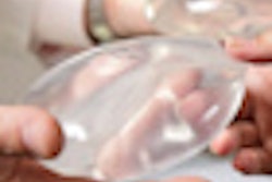
Breast implants were introduced in 1962 by plastic surgeons Thomas Cronin and Frank Gerow, from the University of Texas, U.S., and have evolved rapidly. In the U.K. alone, 10,000 women had implant surgery in 2011, and breast augmentation with single lumen implants remains the most common cosmetic procedure undertaken in the U.K., he stated. Implants also play an important role in breast reconstruction following mastectomy for breast cancer. Given the widespread use of implants, they are often imaged either incidentally as part of screening/cross-sectional investigations, specifically to investigate symptoms/signs of rupture, or when a recurrence is suspected in a reconstructed breast.
In research conducted in Alabama, U.S., for instance, as many as 79% of women with silicone gel implants inserted between 11 to 15 years prior to the study were diagnosed with a rupture on imaging, so it is important that the appropriate modalities are used and findings are interpreted appropriately, Dilks said.
Implants can be categorized broadly by number of lumens, type of filler material, location, and shell material/surface contour. Examples of different types of implants are single lumen, silicone-gel-filled; single lumen silicone gel-saline adjustable, to which can be added a variable amount of saline at the time of placement; single lumen filled with saline, dextran (glucose solution) or PVP (polyvinylpyrrolidone); standard double lumen, silicone gel in the inner compartment and saline in the outer compartment; double lumen, saline in the inner compartment and silicone gel in the outer compartment; adjustable double lumen, silicone gel in both compartments, to which a variable amount of saline can be added in the inner compartment at the time of placement; double lumen, silicone gel in both compartments; and triple lumen, silicone gel in the inner and middle compartments and saline in the outer compartment.
"Although limited in sensitivity in the detection of intracapsular rupture, mammography has a high sensitivity for the detection of extracapsular rupture," he noted. "Abnormality in contour can indicate rupture, although focal herniation may also produce focal contour abnormality without extracapsular rupture."
The sensitivity (56%) and specificity (77%) of ultrasound for the detection of implant rupture make it inferior to MRI, but certain sonographic features can be used to distinguish between gel bleed and intracapsular and extracapsular rupture. However, MRI has been shown to have a sensitivity of 77% and specificity of 94% for the detection of implant rupture. Water, fat and silicone have distinct resonant frequencies, and silicone-only sequences can be performed by suppressing signal from water and fat. Dynamic contrast-enhanced MRI should be performed in cases of suspected malignancy, Dilks recommended.