In the wake of the Poly Implant Prosthèse (PIP) breast implant scandal, the ability to spot the main complication -- a rupture -- is vital, but first radiologists must know what is normal, and that is based on the type of implant the woman has, according to French researchers.
More and more women are getting implants every year, and it's difficult to identify intracapsular and extracapsular ruptures, noted Dr. L-P Berner from the La Croix-Rousse Hospital in Lyon, France, and colleagues, at the recent French radiology congress, Journées Françaises de Radiologie Diagnostique et Interventionnelle (JFR).
In their e-poster presentation, they found single-compartment silicone prostheses are the most common. These implants are filled with silicone gel, and the envelope or "membrane" is silicone elastomer, which is filled with silicone gel or physiological saline. Most silicone implants wear out in about 10 years.
Many women are concerned about their PIP implants following the recent scandal. PIP implants have a porous, more fragile membrane -- they wear out prematurely so the complications are the same but appear sooner.
Patients are monitored using imaging, primarily standard mammography with or without ultrasound. They are usually followed up after one year to ensure there are no complications, and also so the radiologist can become familiar with anatomical changes. After that, however, breast screening is the same as for the general population, according to Berner and colleagues.
The mammography/ultrasound combination is still relevant in first-line assessment, but it's important to pay attention to ultrasound settings, the researchers added. A very normal ultrasound is a very strong predictor of implant integrity.
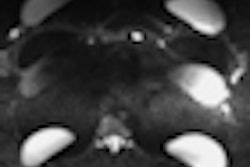
MRI is more sensitive and specific but only for symptomatic patients or patients whose first-line examination indicates a possible rupture. However, MRI is not indicated in the systematic monitoring of women with implants, the researchers noted. MRI should be used primarily for assessing implant integrity and detecting cancerous lesions, especially in women with a history of breast neoplasia. It also has the benefit of a "silicone-only" sequence, as well as two orthogonal planes and a 3D multiplane examination.
Berner and colleagues emphasized the importance of remembering to analyze the armpits and the rest of the mammary gland.
Specifics on ruptures
When identifying ruptures -- using mammography or MRI -- it's important that the modality can indicate whether the rupture is intracapsular or extracapsular, and if there is any siliconoma, especially in the axillary region.
For an intracapsular rupture, no silicone is present outside the fibrous capsule. For an extracapsular rupture, signs of rupture are usually obvious and silicone is outside the fibrous capsule. In extracapsular ruptures, it's important to assess silicone leakage and always specify whether siliconoma is present. If siliconoma is present, identify whether it's single or multiple and the topography.
Also, watch out for false positives, Berner and colleagues advised. Be aware of normal variations in images, including radial folds, and also the patient's history: Implants have may ruptured in the past and been replaced.
On ultrasound, intracapsular rupture appears as echogenic lines, creating a stepladder sign. Extracapsular rupture on ultrasound usually features a snowstorm sign, siliconoma, and axillary silicone adenopathy.
On MRI, if a rupture is present, there will be a linguine sign, subcapsular lines, extracapsular silicone, and siliconoma. If a rupture is possible, other signs must be sought, such as focal detachment of the envelope: the teardrop sign, keyhole sign, or salad oil sign.
Acknowledgment: The original French text of this JFR e-poster was translated by Syntacta Translation & Interpreting, Swindon, U.K.