A computer-assisted surgery system that employs image analysis methods is shaping up well as a tool for orthopedic surgery, Israeli researchers have found.
In a two-part study published online on 9 August by the European Journal of Radiology, the team found that the system, which provides virtual trajectory of the guide wire, intraoperative planning, and various measurements, had a significant positive correlation with manual methods.
"The new computerized software has high reliability in performing measurements of length using an aiming, positioning and referring device intraoperatively," noted a group led by Dr. Eli Sidon of Rabin Medical Center in Petah-Tikva, Israel.
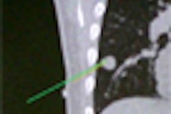
Standard computerized navigation systems suffer from their time-consuming nature; multiple sites are required for inserting the frames of the bone tracker and they must be set up and calibrated for each procedure, according to the authors. As a result, the team sought to evaluate the Surgix guiding system, which combines a set of x-ray-opaque markers that are incorporated into transparent hardware to form an aiming, positioning, and referring (APR) device that's attached to a guide wire.
After surgeons perform fluoroscopy, the images are then processed by the Surgix image processing engine. The APR's 3D orientation relative to the C-arm and drill trajectory in the image is then calculated.
"Since the orientation and trajectory of the APR change with the position of the guide wire, the system can instantly display the guide trajectory or a template of the desired implant as well as accurate 3D measurements on any fluoroscopic image, which includes an APR," the authors wrote.
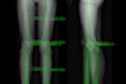
Surgix also provides a panoramic-view image, which is a virtual image that's generated by fusing from 70 to 100 adjacent low-radiation images along the limb, according to the team. Surgeons can utilize this capability for planning osteotomies by measuring length and angulation, placing implants, and reducing long-bone fractures. Image distortions are addressed via a dewarping algorithm.
The researchers sought to evaluate the accuracy of the system in measuring guide-wire lengths and angles as well as measurements via the panoramic-view image tool.
In the first part of the study, the team attached a Surgix APR device to 20 guide wires drilled at different orientations into a synthetic femur model. After the APR device was automatically identified by the system, the guided drill depth was measured. To provide control measurements, two manual measurements were performed using a depth gauge and a caliper.
In the second part of the study, the researchers cut a femur saw bone model in the middle and placed it at various angles. Next, they glued two radiopaque strings of lead balls to both parts of the osteomotized bone to facilitate repeatable measurements. The software was then used to measure 10 panoramic-view images of the saw bone parts that were fixed in different positions to a radiolucent board.
After Pearson correlation coefficient analysis was performed, the researchers discovered a significant positive correlation (r > 0.983, p < 0.01) between all groups of the computerized length and the control measurements. In addition, they found no significant difference among different distances, angles, or positions from the image intensifier.
Furthermore, the panoramic-view image tool and the control measurement had a significant positive linear correlation (r > 0.993, p <0.01) for angle and length measurement.
"The results of the current work validate the accuracy of the measurements of lengths using an APR device and for length and angle measurements on a [panoramic view image]," the authors concluded.