Three-dimensional digital x-ray performs just as well as 3D CT when it comes to measuring the leg-length discrepancies of children and adolescents, according to study published online in European Radiology (19 October 2011). Two-dimensional measurements, however, didn't fare as well.
Leg-length discrepancy in children and adolescents is a common complaint encountered in pediatric orthopedic practices. The discrepancy may be structural or functional. The standard of care is to image with 2D radiography, but it's subject to error from possible torsional deformities and muscle contractures of the ipsilateral or contralateral lower limb. The method also requires careful positioning of the patient.
Microdose digital radiography (DR), CT scanograms, ultrasound, and MRI have all been used to measure leg-length discrepancy in an effort to counter the limitations of 2D x-ray, but the techniques have not been widely adopted and remain unable to assess patients in the functional standing position. Plus, CT requires a high radiation dose.
Another method, biplanar low-dose DR with a 2D/3D system (EOS Imaging), uses two orthogonal sources of radiation and linear digital detectors that produce two orthogonal x-ray images of the lower limbs in the standing position. The system generates surface 3D reconstructions and measurements of the lower limbs, and 2D measurements of the lower limbs, automatically generated from 3D reconstructions.
Until now, no one has determined if there are differences in the 2D and 3D measurements of the lower limbs in children and adolescents, according to a research team led by Dr. Ramon Gheno, from the department of musculoskeletal radiology at Centre Hospitalier, Régional Universitaire de Lille, Hôpital Roger Salengro, in Lille, France.
The group took 3D measurements of eight dried bones, analyzing them with the EOS system using stereoscopic software and comparing the measurements with 3D CT. Also, 47 lower limbs of children and adolescents were studied using biplanar low-dose x-ray 2D and 3D measurements. Both parts of the study were evaluated for femoral length, tibial length, femoral mechanical angle, tibial mechanical angle, frontal and lateral knee angulations, and the femoral neck-shaft angle.
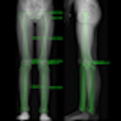
 Above: From left to right, the calibration points, the contours, and the 3D measurements. Below: From left to right, 3D reconstructions views: frontal, lateral, back, up, and down. All images courtesy of Dr. Ramon Gheno.
The researchers found the 3D comparison between the EOS system and CT showed no significant differences in femoral length, tibial length, femoral mechanical angle, tibial mechanical angle, frontal knee angulation, lateral knee angulation, and femoral neck-shaft angle.
However, 2D and 3D measurements from the EOS system demonstrated significant differences in tibial length, femoral mechanical angle, and femoral neck-shaft angle.
"As 3D measurements provide us with more precise information than 2D measurements, we believe that this technique may be a useful and reliable tool in the assessment of lower extremity alignment in children and adolescents, especially when patients present with significant knee flexion/extension or with rotational deformities," the researchers wrote.
The main advantages of the EOS system is its reliability and low radiation dose, which is very important for children, Geno said in an interview with AuntMinnieEurope.com.
"[It also has the] capacity of providing a better parameter for leg limb discrepancies, since 3D measurements are authentic, in contrast to 2D measurements where a 3D structure is visualized in two dimensions," he said.
However, the study has some limitations, mainly the small sample size. Also, the low-dose x-ray semiautomated method of surface 3D reconstruction relies on anatomical landmarks being manually identified, which may lead to more errors than 3D CT reconstructions, the researchers wrote. Lastly, a small number of EOS machines are currently available due to its cost.
Further clinical studies are required -- first, to determine normal 3D values in children and adolescents, and second, to better evaluate young children with partially ossified epiphyses that are still difficult to assess with current software, the researchers wrote.
In his interview, Gheno said the team will present some of the data at this month's RSNA 2011 meeting in Chicago.