Understanding brain development is central to diagnosing congenital malformations that often don't become apparent or are missed in childhood. At the 6th International Symposium on State-of-the-Art Imaging (iSi 2011) in Bordeaux, France, on Thursday, Dr. Birgit Ertl-Wagner urged attendees to improve their knowledge of anatomical development in order to know where to look for abnormalities.
"Many of these disorders are also present in adults, not just in children. The patients don't always go to specialized centers, but to their nearest practice when symptoms appear. Although these cases are relatively rare, pretty much any doctors can be challenged by them," said Ertl-Wagner, section chief of MRI in the Institute of Clinical Radiology at Grosshadern Clinic, University Hospitals of Munich. "In a significant proportion of these cases, the disorders are being missed or misclassified as they are often not well-known."
Typically, imaging of congenital abnormalities takes place in early childhood at the age of 2 to 3 or even, depending on the pervasiveness and severity of the disorder, prior to the child's first birthday. The diagnostic workup is also commonly performed at the beginning of school age, or even later during adolescence and adulthood.
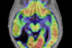
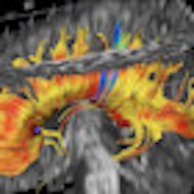
 Diffusion-weighted imaging is proving valuable in cases of suspected ischemia. Diffusion-tensor imaging (DTI) involves fiber tracking and can evaluate the degree of anisotropy and maturation processes. All images courtesy of Dr. Birgit Ertl-Wagner.
The growth in fetal MRI referrals to confirm and classify abnormal findings detected by ultrasound is also increasing diagnosis of prenatal conditions or diseases, as is its capacity to pick up additional abnormalities. Slight dilatation of the ventricles in an ultrasound may reveal severe intraventricular hemorrhaging or absence of the corpus callosum in MRI. This has significant prognostic implications and therapeutic consequences. The imaging expert's role in multidiscipline teams counseling parents on far-reaching decisions, such as termination of pregnancy or potential fetal surgery, should not be underestimated, Ertl-Wagner said.
Besides understanding brain development, the radiologist must look for imaging patterns. Heterotopia is a migration disorder that happens when the neuronal stem cells migrate from the germinal matrix zone to the cortical surface. At some point along the way, they get left behind and don't arrive where they should. In answer to the question posed by a delegate of how to differentiate heterotopia from other white-matter abnormalities, the key factors given were the signal isointensity to cortex and the location along radial migration pathways.
In addition to the signal intensity, structure also provides many vital clues when imaging the brain.
"The signal doesn't need to be abnormal," Ertl-Wagner explained. "With polymicrogyria, for example, where there are too many too small gyri, you usually have a normal signal but an abnormal gray matter-white matter junction."
 Cortex-isointense signal abnormalities in the subcortical white matter on T2-weighted axial sequence corresponding to heterotopia.
Four basic sequences will answer most of the questions aimed at ruling out common malformations in children with developmental delay, Ertl-Wagner told delegates. These are T2-weighted axial sequence, T2-weighted sagittal sequence, FLAIR sequences, and T1-weighted inversion-recovery sequences. Motion correction sequences can be especially helpful when imaging children. In addition, 3D sequences can aid in detecting small abnormalities, in patients with epilepsy, for example.
In terms of field strength, when asked whether 1.5 tesla or 3 tesla was preferable, Ertl-Wagner noted: "3 tesla is especially useful for anatomically small disorders, but one can do really good imaging at 1.5 tesla. It's not only a question of field strength but also of gradient strength and especially of how optimized your sequences are. Sometimes it's more helpful to optimize your sequences than to just go higher in field strength, though ideally it's both."
The usefulness of additional sequences depends on the disorder being imaged. For patients with epilepsy, either adult or child, it may be appropriate to carry out very thin section imaging of the entire brain, such as 3D sequences, with reconstructions through the hippocampi, for example.
This way protocols can be tailored to diagnose the specific reasons why a patient has epilepsy.
 Blurring of the grey matter-white matter interface on an axial FLAIR sequence corresponding to a focal cortical dysplasia in a patient with epilepsy.
The root cause of epilepsy can be very challenging to diagnose as often the abnormalities causing it can be very small, according to Ertl-Wagner. Some of the tiny focal cortical dysplasias with very small signal abnormalities are hard to visualize, and hard to pick up in images of the entire brain, she added.
Here diagnosis requires high resolution, preferably with a very good 3-tesla MR scanner, and optimized sequences, including 3D sequences, such as 3D FLAIR and 3D T1, and then for radiologists to really look for the focus, because finding it or not carries important therapeutic consequences for the patient.
The radiologist must be highly specific about imaging findings, she added.
"Do we find a dysplasia? Do we find a polymicrogyria? Do we find only one or more than one? This influences the therapeutic approach -- for example, whether the focus can be resected or not. If you oversimplify your approach you are going to miss abnormalities and send the patient down the wrong path," she said.