The most common errors in MRI tend to occur in the spine, followed closely by the brain, and then the pelvis, according to a study of MRI mistakes made over seven years at a district general hospital in the U.K.
Dr. Simon Prowse and Dr. Robert Etherington, radiologists at the Countess of Chester Hospital in Chester, reviewed all MRI errors that were confirmed at radiology discrepancy meetings between October 2004 and November 2010. The errors came to light through a systemic review process or by incidental case reviews. When a discrepancy was confirmed by the reviewing radiologists, it was then classified by error type: observation and interpretation (false positive, false negative, and misclassification), technical error, and communication error.
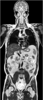 MR investigations of the spine and brain tend to be common sources of errors. Image courtesy of Philips Healthcare.
Of the 99 errors identified in 93 patients, no less than 93 were due to image observation and interpretation mistakes. Of these errors, four were false positives, 68 were false negatives, and 21 were misclassifications. Another three mistakes were due to technical errors, and three more were due to communication errors.
The most common errors by specific investigation involved the spine (27), brain (23), and pelvis (14), according to Prowse and Etherington, who presented their findings at the 2011 U.K. Radiological Conference (UKRC), held in Manchester in June.
The five most common specific recurring errors were failure to recognize, or misclassification of, disk bulge/protrusion/extrusion/sequestration (10 cases); failure to identify distant disease in the pelvis, such as bone lesions (7); failure to identify spondylitic changes in the axial skeleton (6); failure to identify rotator cuff tears (3); and failure to identify orbital disease (3).
"Radiological errors are important to identify to aid patient management and to improve radiological performance," the researchers noted. "By examining seven years' worth of MRI errors, we have identified and presented several recurring errors. We believe awareness of these common errors will help minimize them in the future."
Note: Siemens Healthcare provided the brain MR image used on the home page to illustrate this article.


