The growing popularity of weightlifting has led to a sharp rise in the incidence of musculoskeletal (MSK) injuries, particularly among inexperienced lifters and beginners, Spanish researchers have reported.
"Radiologists play a crucial role in accurately assessing and quickly diagnosing these injuries, with ultrasound and MRI remaining essential diagnostic tools," said Dr. Pablo Vargas Avila and colleagues from Madrid's Hospital Universitario 12 de Octubre. "Lifting injuries are a consequence of one force or a combination of forces, including accidental, overwhelming, tensile, compressive, and overuse or repetitive forces. Strains, tendinitis, and sprains are the most common injury types."
 Fat-suppressed T2-weighted (a,b) and fat-suppressed proton density-weighted (c) axial images. Myotendinous injury at the mid/distal aspect of the insertion of the adductor longus, with hematoma (black asterisks) and surrounding edema (orange arrow), which corresponds to British Athletics Muscle Injury Classification (BAMIC) 2b injury. Follow-up MRI (c) and x-ray (d) depicted a pseudonodular lesion with a surrounding hypointense halo (c) within the previous affected area, related to the evolution of ossifying myositis. All images courtesy of Dr. Pablo Vargas Avila et al, Hospital Universitario 12 de Octubre, Madrid, and presented at ECR 2024.
Weightlifting brings important physical benefits -- including muscular hypertrophy, strength, and endurance -- but also injuries affecting mainly the myotendinous unit, which consists of bone, enthesis, tendon, myotendinous junction (MTJ), and muscle, they told ECR 2024 attendees.
"During weightlifting exercises such as bench press and military press, the rotator cuff undergoes significant stress due to the heavy weights involved. Injuries are more frequently seen in the supraspinatus and infraspinatus," the researchers noted.
The supraspinatus tendon, situated partly in the subacromial space, may experience reduced clearance during overhead movements, and this compression of the tendon can result in tendinopathy. When lowering the barbell from a higher to a lower position, there's rotational stress placed on the infraspinatus muscle, potentially leading to tendinosis, they explained.
"Most weightlifters emphasize training on larger muscle groups, neglecting smaller muscles, creating an imbalance of the internal versus external rotator cuff musculature, rotator cuff-deltoid force couple, and periscapular musculature," they added. "The combination of repetitive loading, unfavorable positioning, and biased exercise selection creates joint and muscle imbalances that increase the lifter's risk of labral tears, labrocapsular junction dysfunction, and shoulder instability."
The elbow and wrist are particularly susceptible to acute injuries when lifting, leading to biceps or triceps ruptures, and chronic overloading may cause overuse syndromes like medial and lateral epicondylitis, ulnar neuropathy, and tendinosis of the biceps and triceps tendons. Most acute injuries of the biceps muscle occur in the distal tendinous insertion, while chronic tears tend to occur proximally, the Madrid team pointed out.
 Ultrasound longitudinal view of distal biceps brachii tendon tear (a). A month later, MRI fat-suppressed proton-density sagittal (b), axial (c), and forearm supinated (FABS) position (d) sequences were performed. Complete full-thickness tear of the distal tendon of the short head of the biceps brachii is visible, with retracted and thickened torn end at the MTJ (white arrow). Some isolated fibers of the partially torn long-head tendon are visible in the radial tuberosity (orange arrow).
The distal biceps tendon is most at risk for acute rupture during eccentric contraction when the lifter weighs 68 kg or more. "This type of motion occurs during biceps curls and rowing movements. Most tears occur 1-2 cm above the radial tuberosity, where there is relative hypovascularity and a histologic transition point."
In the case of a complete rupture, there is a discontinuity with or without retraction, and the proximal tendon is enlarged and demonstrates abnormal signal intensity. If the bicipital aponeurosis is intact, there may be no retraction. The axial view is best for appreciating an intact bicipital aponeurosis, and ultrasound can confirm the tendon's continuity or the abnormal movement of a disconnected proximal tendon, they noted.
 BAMIC 3b injury. Fat-suppressed proton-density weighted axial (a) and coronal images (b) show muscle edema >15 cm (white dotted line) at the myotendinous/intramuscular tendon, with tendon distortion >5 cm (orange dotted line) with a wavy appearance. Interfascial hematoma and surrounding the ischiatic nerve (black arrow). Semitendinous (ST), biceps femoris (BF).
Partial tears involve a change (usually an increase) in the caliber of an abnormal contour of the tendon. Abnormal intratendinous signal intensity is seen on MRI, and on ultrasound, reduced echogenicity, peritendinous fluid edema, bursitis, or hemorrhage may also be visible.
Triceps ruptures are rare injuries to the elbow extensor mechanism that most commonly occur because of a sudden forceful flexion of the elbow against the resistance of a contracting triceps muscle, e.g., during a bench press. "Rupture most commonly occurs at the osseous insertion of the medial or lateral head. It occurs less frequently through the muscle belly or at the MTJ. Complete tears are more common than partial tears."
Hamstring injuries are relatively common in weightlifters, the researchers continued. Competitive lifters exerting maximum effort during the squat or deadlift are at high risk for developing either avulsion of the hamstring tendon insertion on the ischium or a tear of the MTJ.
Gluteal tendinopathy is the most prevalent lower limb tendinopathy. It may be caused by excessive hip adduction, in conjunction with other muscle and bone factors. Complete avulsions of the gluteus medius and minimus from the greater trochanter are unusual, and occurring most often in older women, they said.
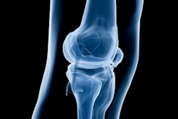
The most common disorder in the leg is tendinosis of the quadriceps and patellar tendons. "Discontinuity of any of the tendinous layers is consistent with a partial tear, whereas transection of all layers is diagnostic of a complete rupture. The patellar tendon ruptures less frequently than the quadriceps tendon. MRI is useful in distinguishing partial from complete tears."
 Quadriceps tendon rupture. Fat-suppressed proton density-weighted coronal (a) and axial (b) images show complete rupture of the conjoined portion of the vastus medialis and lateralis tendons of the quadriceps at its medial margin, with torn end retracted 2 cm (red arrow). Lateral insertion into the patella is preserved (white arrows). Complete rupture of the intermediate vastus (yellow arrow).
In the chest, the usual mechanism of injury to the pectoralis major is the forced abduction of the upper arm that occurs during the eccentric contraction portion of the bench press when the muscle fibers of the lower sternal heads are disproportionately stressed, they concluded.
The co-authors of the ECR 2024 e-poster were Drs. Ana María Bermejo Moriñigo, Andrea Alcalá-Galiano Rubio, Concepción Merino Sánchez, Violeta González Méndez, Cristina Casado Pérez, Andoni Azcona Azcona, Miguel Díez Román, and María Ángeles Antelo Córdov.