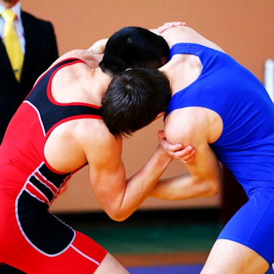
Wrestling is a popular global sport, but due to its arduous nature, it has a particularly high injury rate. Shoulder injuries are a major problem for wrestlers, and new research has shed light on the radiological aspects.
At the first ancient Olympic Games held in Olympia, Greece, in 776 B.C., the main two internationally recognized styles of wrestling were freestyle and Greco-Roman, which are governed by different rules. Greco-Roman wrestlers cannot grab their opponents below the waist.
"Injuries are the biggest nightmare of an athlete," noted Dr. Shalini Agarwal, professor and musculoskeletal radiologist in the department of radiodiagnosis at Pandit Bhagwat Dayal Sharma Post Graduate Institute of Medical Sciences in Rohtak, India.
"Consistent and professional evaluation of the injury pattern sustained by these athletes helps us to understand them and develop a well-designed injury prevention program. Understanding the variables that can cause these injuries helps to minimize the duration of absence from the sport and prolong athletic life," she wrote.
 Tenosynovitis with calcific tendinitis of biceps muscle. Anteroposterior (AP) view of x-ray (top left), coronal fat-suppressed proton density-weighted fast spin-echo MR images (top right and bottom left), and axial CT scan (bottom right) of right shoulder in a 17-year-old wrestler (weight 75 kg, height 165 cm, duration of training three years) shows calcification in the biceps tendon on CT, along with tenosynovitis in this region. The wrestler sustained the injury in competition when in the attack position and was treated conservatively. He returned to practice after 15 days, but he continued to have pain from time to time throughout the study period. All images courtesy of Dr. Shalini Agarwal.
She presented the findings of her study at the 2017 meeting of the European Society of Musculoskeletal Radiology (ESSR). All the wrestlers enrolled in Rohtak's largest wrestling school were included in the study. Over a two-year period from September 2013 to August 2015, the researchers studied 196 wrestlers prospectively with an age range of 9 to 34 (mean age of 19.23 ± 0.28 years), weight range of 38 kg to 120 kg (mean weight of 67.94 ± 1.07 kg), and height range of 123 cm to 195 cm (mean height of 165.62 ± 10.6 cm). A total of 160 engaged in freestyle wrestling, while 36 were Greco-Roman wrestlers.
The participants were of all age groups, weight class, height, and experience. Their shoulder injuries were documented by means of a structured questionnaire, which they completed with help from their trainers. The researchers excluded all old injuries, plus injuries due to systemic diseases, and soft-tissue or bony pathologies unrelated to the practice of wrestling. They performed follow-up until the wrestler returned to practice or competition or quit the sport.
Radiographs were performed either on a digital radiography (DR) system or a computed radiography (CR) system. Image processing or enhancement was applied on DR images and CR images depending on requirement.
Ultrasound was performed using a 5- to 13-MHz linear probe, but in large patients a lower frequency probe of 2 MHz to 6 MHz was necessary. CT was done on a four-slice spiral scanner, and MRI was performed in sagittal, coronal, and axial planes. In all patients, T1- and T2-weighted images were obtained. In addition, T1-weighted gradient-recalled echo (GRE), T2-weighted fast spin-echo (FSE), T2-weighted fat-suppressed, and other sequences, such as proton density-weighted and proton density-weighted fat-suppressed, were done as and when required.
All investigations were performed by a radiologist with 11 years of experience as a general radiologist and one year as a musculoskeletal radiologist. Radiographs were performed in cases of suspected fractures, followed by a CT scan if the diagnosis was in doubt. Ultrasound was performed in all cases of suspected soft-tissue injuries, followed by MRI for further characterization in cases of clinical indication. CT also was performed in cases of suspected osseous injury.
Key findings on injuries
Agarwal and colleagues identified 188 injuries among 121 wrestlers. These injuries were sustained during 36,626 athlete exposures, making an overall injury rate of 5.13 per 1,000 athlete exposures. The case rate was 0.95, while the player rate was 0.61.
After knee injuries, injuries to the shoulder were the most common. A total of 30 wrestlers sustained 35 shoulder injuries (18.6%). The mean age was 18.8 ± 3.22 (range 12 to 27 years), the mean weight was 68.96 kg ± 12.93 (range 42 to 94 kg), and the mean height was 168.33 cm ± 11.225 (range 132.5 to 187.5 cm). Total athlete exposure was 9,743, meaning the injury rate was 3.59, the case rate was 1.16, and the player rate was 0.15.
 Anteroposterior and oblique view of x-ray in a 15-year-old wrestler (weight 94 kg, height 189 cm, duration of training three years) shows a fracture of the surgical neck of humerus (Salter-Harris type I). Injury occurred during training (hard wrestling) in a defensive position when the opponent fell on his shoulder (thunder action). The patient was absolutely normal after six weeks of conservative treatment.
The study authors made the following observations about the shoulder joint:
- 40% of injuries (14/35) were new, while the remaining 60% were recurrent.
- More injuries occurred in competition rather than in practice, and more injuries occurred in the defense position (20/35, 57.1%) than the attack position.
- More injuries occurred in athletes practicing freestyle wrestling (25/35, 71.4%). The injury proportion ratio for the shoulder was 0.7, but this was not statistically significant (p = 0.202)
- There was no statistically significant association with age, weight, height, and duration of practice. Ligament sprains and muscular strains were the most common injuries (37.5%).
- Four patients were advised to undergo surgery, but only one opted for it. Twelve out of 30 wrestlers were absent from practice for less than a week.
Follow-up actions
Agarwal has not done any follow-up studies, but during the study she was able to stress the importance of proper warm-up routines before practice or competitive events and the importance of proper techniques. She was able to teach the wrestlers about the importance of immediate and proper treatment, along with proper physiotherapy and rehabilitation, and the local club acquired new mats and pads, she said.
"There are many problems in my country, and sports medicine is yet to become a separate field of study," she wrote in an email to AuntMinnieEurope.com. "The athletes in my study frequently left after injuries and that is why I had such a large number in the study."
The first phase of her research was a prospective study of knee injuries in wrestlers, published in the Asian Journal of Sports Medicine in September 2016.
The co-author of the shoulder study was Dr. E. Mann from the same institution as Agarwal. For free access to their full e-poster presented at ESSR 2017 via the European Society of Radiology's EPOS database, click here.