Semiautomatic analysis of breast MRI using computer-aided detection (CAD) can provide quantitative data to predict the occurrence of distant metastasis in primary breast cancer, according to a study presented at the 2011 European Congress of Radiology (ECR) in Vienna.
The data could also be used for further risk stratification and in future therapeutic planning for the patients, the researchers concluded.
Prognostic tool
While breast MRI has proved highly sensitive for the detection of breast cancer by providing functional information on tissue vascularization, CAD can semiautomatically contribute quantitative data to assess patient images and limit reader bias.
However, the combination of breast MRI and CAD as a prognostic tool has not been well established due to small studies, said lead study author Dr. Matthias Dietzel from the Institute of Diagnostic and Interventional Radiology at Friedrich Schiller University in Jena, Germany.
The study enrolled 59 consecutive patients with primary breast cancer for one year from January 2004 to December 2004. The researchers excluded patients with secondary breast cancer or other malignancies.
Patients were scanned on a 1.5-tesla MRI system, with utilization of a breast coil and injection of the contrast agent gadopentetate dimeglumine (Magnevist, Bayer HealthCare Pharmaceuticals) before therapy was initiated.
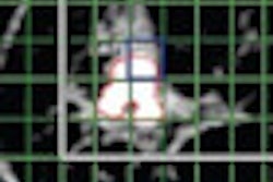
The CAD software and workstation (syngo BreVis, Siemens Healthcare and MeVis) provided semiautomatic quantitative measurement of breast MRI enhancement patterns, which included initial enhancement, washout, peak enhancement, and time-to-peak enhancement.
Clinical follow-up
The standard of reference was clinical follow-up after completion of therapy. Median follow-up time for patients was 1,560 days. "If there was presence of metastasis, it was coded as M1, or if not, it was coded as M0," Dietzel said. "We also did a survival time analysis, so we documented the time interval between the first evidence of cancer in MRI and the follow-up date."
The analysis was able to diagnose six patients with distant metastasis during follow-up with the combination of breast MRI and CAD. Negative predictive value was 100% and sensitivity was 100%.
In addition, washout greater than 40% was identified as significant and an independent predictor for the occurrence of distant metastasis.
"The analysis is exploratory and we could furthermore address more morphological and clinical features, so further evaluation is necessary," Dietzel said. "But we can conclude that CAD and breast MRI using very basic features allows accurate identification of all patients' occurrence of distant metastasis, and these results might be used and implemented in a radiology report for further risk stratification and in the future for therapeutic planning."