Radiology in the Netherlands is in robust shape. This is one of the key conclusions to draw from the list of eight award winners for the 2021 EuroMinnies, which we are proud to reveal. Dutch radiologists won the Most Influential Radiology Researcher and Radiology Rising Star awards, while a Dutch company, Philips Healthcare, collected the prize for Best New Radiology Software.
But other EuroMinnies winners came from across Europe. The Most Effective Radiology Educator award was won by a Spanish radiologist for the second consecutive year, while a Serbian-led European group scooped the Scientific Paper of the Year award for its survey on the use of imaging in the COVID-19 pandemic. Siemens Healthineers collected the Best New Radiology Device, and ImageBiopsy Lab from Vienna received the Best New Radiology Vendor accolade.
In the EuroMinnies award scheme, the full list of semifinalists was based on nominations submitted in late 2020 by members of AuntMinnieEurope.com. The expert panel comprising members of our editorial advisory board, past recipients, and regular columnists selected two finalists to go forward to the finals, and the judges then chose the winners in a second ballot.
Later in March, we will post video interviews with the winners and their trophies so that you can find out more about them and get to know them better.
Most Influential Radiology Researcher
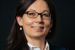
EuroMinnies 2021 winner: Prof. Dr. Marion Smits, PhD, Erasmus MC -- University Medical Centre Rotterdam, The Netherlands
Integrity and an open mind are the two most important personal qualities needed to make a good researcher, according to Prof. Dr. Marion Smits, PhD. She thinks it's absolutely vital to be able to trust colleagues, not only to do their work conscientiously but also to speak up when things aren't going well.
 Prof. Dr. Marion Smits, PhD, from Rotterdam.
"This is crucial for any form of collaboration: Once you know that you can trust your collaborators not to take advantage, you can open up and share your ideas and your data, and create true synergy," she said. "As a researcher you need to be curious, to be open to new ideas but also to alternative explanations, and to be self-critical."
She recalls the moment when neuroradiology and research first gripped her. As a medical student on a neurology rotation, she came across a rapidly deteriorating young patient.
"Nobody knew what was happening to her until the professor of neuroradiology calmly described the abnormalities on her scan, consistent with Creutzfeldt-Jakob disease (CJD). This was at the height of the mad cow disease outbreak, and I was in total awe," Smits explained. "I boldly asked him for a research project, which led to my first publication, and the rest, as they say, is history!"
Her radiology training at Erasmus MC was overseen by Prof. Dr. Gabriel Krestin -- "a better role model in radiology and leadership is hard to imagine, and he has continued to mentor me," she noted.
An important figure in her academic career at Erasmus MC has been Prof. Dr. Myriam Hunink, PhD, who was Smits' PhD supervisor. "I just love how she is a very mathematically oriented researcher on the one hand, but also has such an eye for 'soft skills' -- well before this became trendy. She has taught me some really important skills on time management and self-care."
Smits said her greatest inspiration is her own research group -- from the bachelor student to the assistant professor. "Seeing how they look at the world, tackle problems, are innovative, follow their dreams, and develop in their own careers. That is really the highlight of my work."
Formally, 40% of her time is spent on clinical work and 60% on research and management responsibilities, but she admits it does sometimes feel like having two full-time jobs. She tries to protect her clinical time by not having any meetings planned on those days and being fully focused on patient care. Her PhD students are her highest priority, though.
Smits said it helps that she's well organized, using Mailbutler to manage her emails and Evernote to keep track of tasks, but doing research in a clinical environment is still hard work that often must be done in one's spare time.
"It's really important to work on a topic and with people that you like," she commented. "However motivated and smart you are, if you're not supported properly then it's going to cost you so much energy, and that's just going to make it unbearably hard and frustrating. Don't be afraid to say no to a project or a supervisor that really doesn't fit."
Look for research projects that are well-defined, and make sure you find out from the start what's expected from you and what you will get out of it, she recommended. If you can't find this in your own institution, look at opportunities elsewhere, she added.
Getting funding for research is terribly hard, and Smits doubts she's found the best approach yet. She tries not to get too upset if an application gets rejected and celebrates when she gets lucky. Also, when writing an application, she focuses on the creative aspect of putting a new research idea together and does not worry too much about the outcome.
The pandemic has had a huge impact on her work in the past year. During the first wave of COVID-19, all projects were put on hold, and all researchers were banned from the hospital.
"I found the early months of the pandemic very difficult, as I felt very responsible for the wellbeing of all the people in my research group, some of whom were on an exchange and/or from abroad and cut off from their families. At the same time, I had to guide my clinical neuroradiology group," she said.
Smits started a chat group and online coffee breaks, which has kept the group together, but she remains concerned about the isolation and loneliness some of these young people face. There are still many restrictions on research projects, so she is worried about delays in completing projects and PhDs. On the positive side, it's been great to be able to invite many external speakers to online meetings, she said.
At ECR 2021, she will give several invited lectures as well as a scientific presentation, the latter on the impact that the pandemic has had on the professional life and personal wellbeing of the neuroradiology community. She's also moderating a session on ultrahigh and ultralow-field MRI and doing a Table Talk about her work as chair of the Publications Committee of ESR.
Outside of radiology, she loves running, and she particularly enjoyed doing the half marathon on a gloriously sunny autumn Saturday afternoon in Oslo, when a congress coincided with the city's marathon weekend.
"Life doesn't get much better than that!" she declared.
You can read more about her research activities on her personal website.
Runner-up: Prof. Dr. Marc Dewey, Charité, Berlin
Most Effective Radiology Educator
EuroMinnies 2021 winner: Dr. Luis Martí-Bonmatí, La Fe University, Valencia, Spain
"Education is based on leadership and communication. The educator has to be honest, synthesize relevant aspects to understand the imaging expression of the disease, use visual schemes for those features to be highlighted, provide an innovative view on how to improve daily radiological efficiency, foster a multidisciplinary collaboration, and be open to the opportunity to learn from the discussions," Dr. Luis Martí-Bonmatí told AuntMinnieEurope.com.
 Dr. Luis Martí-Bonmatí from Valencia
His advice to other educators is to construct useful, evidence-based, innovative, and easy to recognize messages, despite the inherent complexity of our specialty.
"Keep it as simple as possible, keep it updated, keep it attractive and precise. Be modest and interactive. And practice, as experience is the mother of teachers," he said. "We learn by listening to others, with a willingness to understand, and by critical thinking. We should question what we can learn that is important to healthcare, which aspects should be openly discussed. And then, work and teach on them."
Martí-Bonmatí started out by learning and teaching clinical radiology. He enjoyed the basics of MRI, liver tumors, and perfusion studies with contrast media. From there, he jumped to dynamic parameters of diffusion and perfusion studies, and seamlessly to imaging biomarkers, structured reports, and artificial intelligence (AI).
"I have always been excited about how to improve radiology -- learning its biological bases, promoting a closer relationship to the real needs of patients. If you like clinical radiology based on evidence and research, then you can share and spread this interest and knowledge, even between more classical and less innovative colleagues," he noted.
His mentors in radiology have included colleagues at the European Society of Gastrointestinal and Abdominal Radiology (ESGAR) and the European School of Radiology (ESOR), from whom he has learned at the summer school and visiting scholarship programs. He was inspired by Prof. Nick Gourtsoyiannis for his ability to stir the imagination, Prof. Adrian Dixon and Prof. Dr. Max Reiser for their academic leadership, and Dr. Bruce Hillman for his strategic scientific vision.
At ECR 2021, Martí-Bonmatí will moderate two sessions (AI in unmet clinical needs; and radiology in the cloud: advantages and threats). He will also give a lecture on the ESOR Coffee & Talk Sessions on "10 tips & tricks for structured reporting," and one other on-demand lecture titled "How far are we in getting AI into clinical practice?"
COVID-19 has hit Spain particularly hard.
"The pandemic has shaken society, killing people and devastating economies. Our lives are completely different now," he said. "Teaching is either online and quite distant or frozen. My feeling is that teaching must involve face-to-face activity. On-demand videos, podcasts, and live discussions are attractive and work in some way, but cannot be a substitute for direct interaction with the audience. Mixed face-to-face and online courses might be a chance to improve dissemination."
In spite of this environment, an educator's daily activity can be very entertaining if you enjoy your work.
"Running a department, a research group, and a prestigious journal can be very tedious if you do not have a team of enthusiastic and collaborative people. Fortunately, I have the support of all radiology subspecialty leaders in my hospital duties, of my colleague Angel Alberich and Ana Penades in the research group, and of Stefanie Muzik and Irene Christoffel at the Insights Editorial Office. And of course, the love of my wife Cristina and my sons at home and of my friends.
"All of these activities fill the day, but we do still have the night!" he stated.
Outside of radiology, he likes reading books on literature and general science, listening to music, strolling with his dog, enjoying good wine, walking the Camino de Santiago with close friends, watching movies with his wife, and making his family happy.
"And doing this with calm and no rush," he concluded.
Runner-up: Dr. Adrian Brady, Mercy University Hospital, Cork, Ireland
Radiology Rising Star
EuroMinnies 2021 winner: Dr. Merel Huisman, PhD, University Medical Center Utrecht, the Netherlands
Many radiology trainees have found life particularly hard during the COVID-19 pandemic, but Dr. Merel Huisman, PhD, a final-year radiology resident, has some advice based on her own experiences.
 Dr. Merel Huisman, PhD, from Utrecht.
"Listen to your inner voice, be yourself, and find what truly brings you joy professionally," she said. "Stop doing career-related things just for your curriculum vitae or the labor market, because we never know what the future holds. Instead, keep a clear vision of your strongest skills and don't be afraid to use them, even in a junior position. This way you'll be able to move mountains while having fun and connecting with people around you."
Huisman has a keen personal interest in the intersection between AI and clinical epidemiology, alongside her other subspecialty interests of cardiothoracic and musculoskeletal imaging. She was the key investigator in an international survey on change management of AI in radiology, which she conducted with her former colleague and friend Dr. Martin Willemink, PhD.
"Change management has always been one of my most favorite topics. We thought this would be a quick project but, as always in research, it was more work than anticipated with over 1,000 responses in multiple languages, compounded by the fact that I had to do it all in my free time in addition to my residency, and on top of many other extracurricular activities and quite an active social life prepandemic," she told AuntMinnieEurope.com. "It resulted in three oral presentations at ECR 2020 and two papers that were almost immediately accepted by European Radiology."
In addition, in 2020 she spoke about AI in radiology at the Dutch Society of Radiology (NVvR) annual meeting and a meeting of the National Institute for Public Health and the Environment (RIVM). In December 2020, she became a trainee member of the editorial board of Radiology: Artificial Intelligence, while in January 2021, she became a junior board member of the Thoracic Radiology Section of the NVvR. She also serves as a scientific reviewer on the imaging informatics subcommittee for ECR 2022.
Huisman cites several key figures who have helped shape her career, two of whom she met in her final year of medical training: Prof. Dr. Maurice van den Bosch, PhD (UMC Utrecht at the time, now CEO of OLVG Amsterdam), who helped to hone her leadership skills and professional development activities, as well as Prof. Dr. Lenny H.M. Verkooijen, PhD, the co-supervisor on her PhD in interventional radiology.
During this four-year period, Huisman learned the fundamentals of good translational research in a high quality research environment.
Also, Prof. Dr. Tim Leiner, PhD, inspired her in 2018 to attend a symposium on AI in radiology in Nijmegen. She decided to dedicate herself to improving the responsible use of AI in imaging while promoting the involvement of women in these more technical topics. At that same conference, she met Dr. Paul Algra, who asked her to join the editorial board of MemoRad, the official journal of the Radiological Society of the Netherlands, and Dr. Erik Ranschaert, PhD, who in 2019 asked her to join the European Society of Medical Imaging Informatics (EuSoMII) as a board member. Following this, she became chair and founder of the EuSoMII Young Club.
"These roles have allowed me to learn skills that go beyond my residency program," noted Huisman, who since 2018 has also been chief radiology resident at Meander Medical Center in Amersfoort.
Another inspiring colleague has been Prof. Dr. Cornelia Schaefer-Prokop, PhD. Additionally, working abroad has contributed to her personal and professional development.
"I've been incredibly fortunate to have had the opportunity to study and work abroad three times: in Pondicherry in India, in Paris, and in Dallas. These experiences have made me very independent, resilient, resourceful, and grateful," she said.
Runner-up: Dr. Samantha Fossey, Royal Sussex County Hospital, Brighton, U.K.
Most Significant News Event in European Radiology
EuroMinnies 2021 winner: ECR 2020 moves fully online for first time
When the COVID-19 outbreak began to spread in northern Italy during the second half of February, the European medical imaging community faced an agonizing wait. Would ECR 2020 begin onsite in Vienna on 3 March, as planned?
After much deliberation, the ESR postponed the congress eight days before it was due to start, and it rescheduled the meeting to 15-19 July. The ESR considered taking legal action against the City of Vienna and the Republic of Austria due to the authorities' alleged failure to provide clear guidance and direction over whether to call off ECR 2020, but it did not sue in the end. On 9 April, the ESR announced that the congress would be held online-only, not onsite.
"It is with a heavy heart and much regret that the Board of Directors of the ESR has decided not to hold an onsite ECR in 2020," the ESR statement noted. "We have a social responsibility to our members, partners, staff, and the general public. In the current crisis, the ESR must lead by example, and it is clear that your safety will always be our No. 1 priority and we sincerely hope for your understanding and support of our decision."
Online conferences must find new ways to allow discussions involving remote participants during all sessions to create the sense of community and collective learning felt at onsite events, according to a follow-up statement issued on 16 May by the ESR. To remain relevant, scientific societies must adapt to this new reality.
"This will not be easy, but making this necessary shift successfully will establish a new standard for what defines medical conference success in the future," the ESR Board of Directors noted. "The COVID-19 pandemic has changed how humans interact with each other, and these changes are likely to persist, at least for some years."
The pandemic has had an effect on every aspect of life across the globe during the past year, but its impact has been felt particularly acutely by conference organizers such as ECR and RSNA. Huge uncertainty still surrounds the future of congresses, and the prospects of any large face-to-face meetings taking place in 2021 are rapidly diminishing.
Runner-up: Discussion in the Netherlands over AI replacing radiologists
Scientific Paper of the Year
EuroMinnies 2021 winner: The use of imaging in COVID-19 -- results of a global survey by the International Society of Radiology. Blažić I et al, European Radiology, 17 September 2020
Tennis legend Novak Djokovic has made a series of controversial comments about COVID-19 over recent months, but he is not the only Serb to have made the headlines across the globe. In a much more calm and balanced way, Dr. Ivana Blažić, PhD, MRI section head in Clinical Hospital Center Zemun in Belgrade, has also made a significant contribution and impact during the pandemic.
 Dr. Ivana Blažić, PhD.
She led a group of authors last year that collected information regarding current imaging practices in the management of patients with COVID-19 depending on the severity of disease and particular clinical scenarios. Their survey of 50 radiology departments around the world conducted by the International Society of Radiology is the winner of the Scientific Paper of the Year.
Blažić's team discovered that the COVID-19 pandemic significantly impacted the imaging department's routine activity. Most institutions used chest imaging in suspected or confirmed patients with COVID-19 showing severe symptoms, although 69% tended not to use imaging in asymptomatic patients. However, the group found noteworthy variations in how imaging was used for COVID-19 patients for different disease severity and various clinical scenarios in regard to imaging modality type used.
The team plans to present results from this study at the upcoming ECR 2021 EuroSafe Imaging Poster Exhibition, Blažić said.
"The majority of patients with suspected or confirmed COVID-19 initially undergo chest radiography, although some may need chest CT or lung ultrasound, depending on disease severity and information needed for clinical decisions important in patient management," she told AuntMinnieEurope.com. "Additionally, other modalities, including conventional radiography, ultrasound, CT, and MRI, have their roles in response to out-of-chest COVID-19 presentations or for any other medical condition in patients with the disease."
Blažić and colleagues hope the survey results will help to understand differences in the organization of radiology departments around the world and to fill knowledge and care gaps in how these departments handle the COVID-19 pandemic by fostering the development of an overall strategy for radiology department organization and imaging protocols in pandemic conditions.
In fact, Blažić is a member of the ISR's Communications Working Group and has served as a lead writer of the World Health Organization rapid advice guide for the use of chest imaging in COVID-19. This guide, developed by an international expert panel based on a systematic review of the scientific evidence, was also informed by this research.
"Those recommendations may help in global standardization of the use of chest imaging in patients with COVID-19, since they cover complete clinical pathway of COVID-19 patients, paying specific attention on different resource settings and healthcare organization levels in each member state," she said.
Runner-up: Mortality due to cancer treatment delay: Systematic review and meta-analysis. Hanna T et al, BMJ, 4 November 2020.
Best New Radiology Device
EuroMinnies 2021 winner: Magnetom Free.Max MRI scanner, Siemens Healthineers
For years, it's been thought that high-field scanners won the struggle over whether low-field or high-field MRI would predominate in radiology. But the winner of this year's EuroMinnies award for Best New Radiology Device, Magnetom Free.Max, flips the script of that debate, in the process offering a vision of what MRI might have been -- and what it might still become.
 The first installed Magnetom Free.Max in the children's clinic at the University Hospital Erlangen in Germany. Image courtesy of Siemens Healthineers.
Magnetom Free.Max is a superconducting 0.55-tesla scanner that Siemens has designed specifically to go places where MRI isn't usually found. The company announced the system in November 2020, and it plans to start commercial shipments later this year.
Siemens Healthineers is promoting Free.Max as the smallest and most lightweight whole-body scanner the company has ever brought to market, while also being very economical to operate. The company is also highlighting the clinical versatility possible with the system's wide 80-cm bore opening.
The vendor believes that there is untapped potential to place MRI at locations -- like intensive care units and even urgent care centers -- where it hasn't previously been economical due to the size of a typical superconducting MRI scanner and the cost of installation. These locations typically use CT instead.
But MRI has a range of benefits not available with CT, such as detailed soft-tissue resolution and a lack of ionizing radiation. With respect to Free.Max, the scanner only requires 1 liter of helium to operate, with no refills. This means the system can be installed without the need for a quench pipe.
The scanner's 80-cm bore makes it ideal for scanning claustrophobic or obese patients, and the system has a table weight limit of 320 kilos. It includes technology like the company's myExam Companion to assist radiographers in setting up the system.
The first Magnetom Free.Max has been in operation at a children's clinic in University Hospital Erlangen in Germany, where studies comparing the system's image quality to that of a 1.5-tesla scanner have produced excellent results, according to Arthur Kaindl, PhD, head of MR at Siemens.
"The old paradigm of field strength leading to image quality is gone," Kaindl said. "The first radiologist said, 'I can't believe how good the images are.' "
What's more, researchers there are finding that it's more convenient to scan patients in the pediatric department rather than send them to radiology -- an important consideration during the COVID-19 pandemic.
A second Magnetom Free.Max is currently being installed in the radiology department at the University Hospital of Basel in Switzerland, and it may be used in the emergency room and intensive care unit in the future. Kaindl told AuntMinnieEurope.com that a single engineer was able to maneuver the system through the hospital's doorways and rig it himself. Siemens believes that Free.Max's size and weight will reduce installation costs by several orders of magnitude, he said.
Siemens expects to receive the CE Mark for Magnetom Free.Max in April, with official shipments to begin in August, Kaindl said. The company also plans to submit the scanner for regulatory approval in the U.S.
Runner-up: Aquilion Exceed LB CT scanner, Canon Medical Systems
Best New Radiology Software
EuroMinnies 2021 winner: Advanced Visualization Workspace -- IntelliSpace Portal 12, Philips Healthcare
Philips Healthcare has won its second straight EuroMinnie award in the category of Best New Radiology Software, claiming this year's trophy for its Advanced Visualization Workspace - IntelliSpace Portal 12 software.
Introduced at RSNA 2020, the latest version of Philips' advanced visualization platform features a wide range of new quantitative imaging and workflow features, including the most artificial intelligence (AI) enhancements of any product in the company's portfolio, according to Calum Cunningham, general manager of enterprise diagnostic imaging at Philips.
 Philips has included new AI algorithms on IntelliSpace Portal 12 designed to detect lung nodules, analyze cardiac function, and quantify pulmonary infiltrates. Image courtesy of Philips Healthcare.
"We have also taken steps to provide more automation to current workflows and faster time to achieve their desired results than ever before," Cunningham told AuntMinnieEurope.com.
IntelliSpace Portal 12 includes several new AI-powered clinical software packages to support quantitative image analysis in cardiology, neurology, oncology, and pulmonology. In cardiology, for example, Philips' MR Cardiac Analysis application provides AI-based pre-processed ventricle segmentation, while the vendor's MR Caas 4D Flow Analysis software aims to make quantifying blood flow patterns in the heart and main arteries faster and more reliable, according to the company.
Another new AI package, CT Pulmo Auto Results, provides automated and quantified assessment in COVID-19 patients. IntelliSpace Portal 12 also now supports automatic CT brain perfusion analysis; these measurements are instantly available in PACS and can be emailed to clinicians within two minutes, Philips said. Other quantitative imaging and workflow capabilities include multimodality tumor tracking, as well as AI-based detection, characterization, and tracking of lung nodules over time.
In addition, IntelliSpace Portal 12 features the company's Photo Realistic Volume Rendering (PRVR) technology, which was designed to enable images to be viewed as true to "real life" as possible and support the use of radiology images as an educational and communication tool.
"And you have the flexibility to manipulate light and shadow on anatomical structures for better understanding of depth and spatial relation between key anatomical structures, as if you'd be looking at them in real life," Cunningham said. "It can be optimized for different anatomies from cardiac and vascular to pulmonary and MSK."
Philips has integrated IntelliSpace Portal 12 with its Vue PACS software, enabling the platform's advanced visualization applications to be accessible within the PACS client during interpretations, stated the vendor.
"IntelliSpace Portal's automated results are integrated within the primary reading workflows with Vue's interactive reporting system both with key images, quantified data, and graphs," Cunningham said.
Runner-up: BerlinCaseViewer app for COVID-19 case recognition, BerlinFlame GmbH
Best New Radiology Vendor
EuroMinnies 2021 winner: ImageBiopsy Lab
If at first you don't succeed, try, try, try again. This adage certainly applies to ImageBiopsy Lab, which develops AI-based software for the diagnosis of musculoskeletal (MSK) diseases.
"The main difficulty, as for any startup, is to secure sufficient funding to get your idea off the ground. For us, this was initially end of 2016 when we applied for a significant startup fund in Austria called aws-Seed," explained CEO and co-founder Dr. Richard Ljuhar.
 The ImageBiopsy Lab management team. From left to right: Christoph Goetz (chief operating officer), Philip Meier (chief commercial officer), Richard Ljuhar (CEO), Matthew DiFranco (chief scientific officer), Zsolt Bertalan (chief technology officer).
While the company's initial pitch failed, the team got a second chance in mid-2017. With a reworked business plan, it convinced investors of the company's technology and go-to-market strategy.
"This experience was extremely valuable for us as it is easy to get lost in your own ideas, so having this pressure to compete with other innovative startups helped to shape the focus and taught us how to flexibly adapt to customer feedback," Ljuhar told AuntMinnieEurope.com.
He offers the following five tips to any digital health startup:
- Listen to your customers, especially the paying ones. It's often easy to convince a potential customer to use an AI software free of charge, but once medical professionals are willing to pay for it, this is when they see real value in it. So efficiently addressing the pain point and translating it into the right tools is critical.
- For AI software, adding any additional step to the workflow is a no go. Ensuring seamless integration is key.
- As the company grows, the organizational structure becomes increasingly important. Scaling up will become a nightmare if you haven't started working on this issue prior to pushing sales and the product pipeline.
- Go out to the market as early as possible, even if you just have a minimum viable product, especially if you come from an academic background.
- Build early on a strong key team, with members that trust and support each other
Being in the field of digital health, the company has been able to keep the team and customers engaged during the pandemic. Furthermore, its chief software architect moved the entire IT infrastructure into the cloud, so this gave the team the option of working remotely without any interruptions.
Another benefit of being in digital health is how the company deploys its software and interacts with customers: ImageBiopsy Lab's MSK solutions are deployed completely remotely, and it switched most of its sales campaigns to virtual interactions.
"The face-to-face meeting can't be replaced by video conferencing indefinitely," Ljuhar continued. "Consequently, we have high hopes for the remainder of the year that we can travel again - especially to our team in the U.S."
ImageBiopsy Lab's AI software applications such as KOALA (knee osteoarthritis labeling assistant), PANDA (pediatric skeletal maturity assessment and growth potential estimation), and HIPPO (femoroacetabular impingement and hip dysplasia) assist in the diagnosis of MSK diseases, such as knee osteoarthritis, leg-length discrepancy, and femoroacetabular impingement.
The company's applications can save up to three minutes of interpretation time per x-ray. This can add up to 45-60 extra minutes per day in time savings for a radiologist.
In November 2020, ImageBiopsy Lab signed a deal with GE Healthcare in which its software will become a part of the Edison AI ecosystem that GE is building. It also signed a deal with Siemens Healthineers in July 2020 in which its KOALA software will be made available to Siemens customers.
At this year's ECR Online the company will focus mainly on its scientific presentations and presence at its partners' booths such as Siemens Healthineers and GE.
Runner-up: Deepspin, Berlin, Germany