Italian researchers have used augmented reality (AR) technology to overlay dynamic virtual 3D models based on MRI scans directly onto patient anatomy. In a recent article in European Urology, they found their method to be highly effective for prostate cancer surgery.
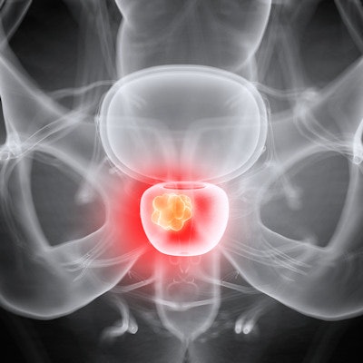
Prostatectomy is often necessary for cancer patients who have tumors breaching the prostate capsule, or external protective layer. Intraoperative guidance plays a crucial role in helping clinicians identify such tumors during surgery. However, conventional 2D image guidance relies on modalities such as MRI that offer a limited visualization of patient anatomy, noted first author Dr. Francesco Porpiglia and colleagues from the University of Turin.
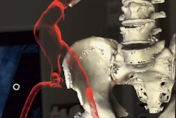
In a previous preliminary trial, the researchers demonstrated a new proprietary technique for generating highly detailed and elastic virtual 3D models of the prostate based on multiparametric MRI scans. These 3D models not only capture the structure of the prostate and tumors encroaching upon the prostate capsule but also take into account the organ's elasticity and susceptibility to deformation during surgery.
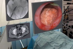
For the current study, the group integrated AR software into the computer of a robotic da Vinci surgical console. Then clinicians used the technology to overlay patient-specific virtual 3D models of the prostate and nearby tumors onto an endoscopic view of the actual structures in real-time (Eur Urol, 9 April 2019).
The researchers evaluated the ability of the surgeon to complete the nerve-sparing phase of robot-assisted radical prostatectomy for 20 patients with prostate cancer using their AR-based technique. For comparison, they also performed the same procedure for 20 different prostate cancer patients using a standard protocol involving multiparametric MRI guidance.
They found that the surgeon was able to identify the precise location of cancers that were invading the prostate capsule much more accurately by using the group's AR technique than by examining multiparametric MRI scans. On average, this improvement allowed the surgeon to partially spare more than double the number of nerves during surgery with the AR technique than with the standard method.
| AR vs. multiparametric MRI for guiding robot-assisted radical prostatectomy | ||
| Multiparametric MRI | Augmented reality | |
| Accurate identification of cancer invasion in prostate capsule* | 47% | 100% |
| Partial nerve sparing* | 35% of nerves | 85% of nerves |
| Mean operation time | 117.5 minutes | 140 minutes |
| Median hospital length of stay | 5.6 days | 5.3 days |
| Continence recovery 1 month after surgery | 85% of patients | 90% of patients |
Ultimately, they showed that examining the elastic 3D virtual models with AR technology facilitated robot-assisted radical prostatectomy and demonstrated the potential to improve functional outcomes, compared with conventional methods.
A major limitation of the technique is the long amount of time it takes to complete manual image segmentation of the prostate and surrounding structures, the authors noted. The intricacy of the invasive prostate cancer observed in this study required an experienced radiologist and urologist to work together to segment the images.
Still, implementing 3D imaging and AR into cancer surgery appears very promising, with growing interest among clinicians to use the technology for other procedures such as partial nephrectomy, they added.
"This new implementation of the robotic platform can represent a new paradigm of the treatment of [prostate cancer] in the 'precision surgery' era," the authors wrote. "The procedures can be tridimensionally modulated in real-time on the basis of a patient's specific anatomy and, in particular, [capsular involvement] location."