A wording problem exists in the current debate about gadolinium deposition, and it's essential for radiologists to become better informed about the newest animal studies and the underlying theory of gadolinium deposition, according to Dr. Alexander Radbruch, JD, a leading German researcher in the field.
"From a chemical and toxicological point of view, it is a huge difference if the intact complex is temporarily in the brain -- and possibly washed out over time -- or if the gadolinium is released from the complex and binds to other partners. However, both events are often referred to as gadolinium deposition," he told AuntMinnieEurope.com.
 Dr. Alexander Radbruch from the German Cancer Research Center in Heidelberg and the University Clinic Essen.
The vital point is to focus on the kinetic character of the gadolinium deposition, explained Radbruch, who is an associate professor of radiology and neuroradiology at the German Cancer Research Center in Heidelberg and the University Clinic Essen.
After the initial paper by Murata and colleagues that showed gadolinium after injections of macrocyclics -- and did not differentiate between chelated and dechelated gadolinium -- in the brain tissue of deceased patients, it was reported many times that macrocyclic gadolinium-based contrast agents (GBCAs) also deposit in the brain, he added. However, given the results from the latest study led by Philippe Robert, PhD, from the department of research and innovation for the Guerbet Group and published on 22 May in Radiology, it might be the case that the intact macrocyclic chelate was in the washout phase and that no gadolinium (or at least much less gadolinium) would have been found at a later point in time, he continued.
"We have to differentiate the propensity of a GBCA to dechelate in vivo and its clearance. Both parameters are unfortunately often mixed up in the debate," Radbruch noted. "If we compare the amount of retained total gadolinium shortly after injection (e.g., one week, as in the McDonald animal study), potentially varying clearances of the GBCAs are a relevant factor for the remaining total gadolinium. If we wait for a longer period (e.g., half a year or a year), the varying dechelation of the agents will determine the total amount of retained gadolinium much more than the clearance."
Research on other organs
The Robert study focused only on the brain, Radbruch noted, whereas his own group is conducting studies in which other organs, such as the heart, skin, bone, and liver in animals and in a patient's tissue, are assessed. He hopes they will soon have more evidence about whether the proposed mechanism of gadolinium deposition in the brain is similar in other tissues and, more importantly, whether the different forms of gadolinium deposition are correlated with any pathological changes.
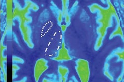
 Images of tissue of the human cerebellum: On the left is gadolinium, and on the right is phosphor. Gadolinium deposition in the dentate nucleus of a patient who received the linear nonionic GBCA gadodiamide (injections of other GBCAs cannot be excluded). Accumulation of gadolinium displays within the tissue of the dentate nucleus. The structure of the tissue is visualized by displaying phosphor on the right side. Image courtesy of Prof. Uwe Kartst and Stefanie Fingerhut, PhD, University of Münster.
The difference between linear and macrocyclic GBCAs is based on how the ion of gadolinium binds to a ligand to create the contrast agent. The bonds also allow the contrast agent to be expelled from the body after an MRI scan with no residual gadolinium left behind in a process known as chelation. However, some researchers postulate that the connection somehow breaks down in linear GBCAs through dechelation and allows gadolinium to remain in patients.
"The current study (from Robert et al) provides further evidence that gadolinium deposition in the brain needs to be specified as either deposition of the intact chelate that is eliminated over time for both linear and macrocyclic GBCAs, or as potentially permanent dechelated gadolinium deposition that is caused exclusively by linear GBCAs," Radbruch wrote in an accompany commentary in Radiology (22 May 2018).
The study investigates only one linear GBCA and one macrocyclic GBCA, and conclusions should be drawn individually for all GBCAs in use, Radbruch warned.
"At the current stage of the debate, it is extremely important to emphasize that despite the enormous number of GBCA injections, no clinical correlates are known and that any unreasonable decline of GBCAs in clinically indicated situations should be avoided," he added. "Nevertheless, we should remind ourselves that chelated GBCAs are injected because we thought they would be excreted over time and not dechelate in vivo."
Paris in June
Radbruch will be presenting in Vancouver, British Columbia, at this week's American Society of Neuroradiology's annual congress and the joint meeting of the International Society for Magnetic Resonance in Medicine and the European Society for Magnetic Resonance in Medicine and Biology, which begins on 16 June in Paris, but he does not think any significant new results will be ready in time for the Paris congress. He said he is very optimistic that we will see new studies during this year.
The study by Robert and colleagues was performed in mice and found that the amount of gadolinium retained in the brain after the administration of a GBCA depends heavily on whether the agent was linear or macrocyclic. They observed that as much as 75% of the total gadolinium detected in the cerebellum after the injection period is retained one year after the last injection of gadodiamide (Omniscan, GE Healthcare), a linear agent. By contrast, the researchers found no gadolinium above the level of detection, with macrocyclic GBCA gadoterate (Dotarem, Guerbet).
"Further investigations are needed to characterize the nature of these macromolecules and to provide a better understanding of the putative risk associated with this gadolinium retention in the brain," the researchers wrote.