Children with migraine headaches have a characteristic pattern of gray-matter changes that may be useful as a biomarker of the condition, according to Italian investigators who presented their findings at the American Academy of Neurology (AAN) 2013 annual meeting in San Diego.
The team of neuroradiologists and neuroscientists headed by Dr. Massimo Filippi of San Raffaele Scientific Institute and Vita-Salute San Raffaele University in Milan found that children with migraines had significant gray-matter atrophy of the front and temporal lobes in comparison with controls. They also had significantly higher gray-matter volume in the right putamen.
The researchers examined MR images from seven children who had migraine with aura and another five who had migraine without aura. Their goal was to extend work in adult migraineurs that showed structural brain abnormalities are not correlated with patients' clinical characteristics. This had suggested the structural changes were not caused by damage to the brain during repeated migraine attacks but, rather, could serve as an independent marker to confirm the presence of the disease.
The Italian subjects' average age was 14 years, half were female and their average disease duration was 3.7 years. The mean attack frequency was seven per year for the children with aura and 23 per year for those without aura. The researchers compared imaging data from these children with those from 15 age- and sex-matched healthy controls who did not have a family history of migraine, a personal history of migraine, or any other neurological dysfunction.
Three types of images were acquired from each subject: 3D T1-weighted fast field echo, T2-weighted turbo spin echo, and fast fluid-attenuated inversion-recovery (FLAIR).
Four of the patients without aura had small, punctate T2 hyperintense lesions at the level of the subcortical and periventricular white matter. The researchers discovered the migraine patients had significant gray-matter atrophy of the frontal and temporal lobes compared with controls -- specifically, in the left inferior frontal gyrus, the left middle temporal gyrus, the right orbitofrontal gyrus, and the right inferior temporal gyrus.
They also found the pediatric migraineurs had a significantly larger volume of gray matter in the right putamen than did the controls.
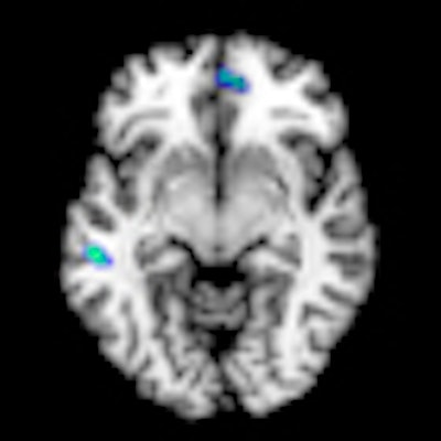
 Areas showing significant gray-matter (GM) volume differences (p < 0.001, uncorrected) between migraine patients and healthy controls, superimposed on a high-resolution T1-weighted template. Areas with reduced gray-matter volume in migraine patients versus controls are shown in blue; areas with increased gray-matter volume in migraine patients versus controls are shown in red. All images courtesy of Dr. Massimo Filippi.
In other findings, the researchers found migraine patients with aura had significantly higher gray-matter volume in the left fusiform gyrus than those without aura.
 Areas showing significant GM volume differences (p < 0.05, familywise error corrected) between migraine patients with (MWA) and without (MWoA) aura, superimposed on a high-resolution T1-weighted template. MWA vs. MWoA: GM volume increase of the left fusiform gyrus.
There were not any significant white-matter changes in migraine patients compared with nonmigraine patients, nor was there a correlation between gray-matter abnormalities and disease duration or attack frequency.
"The absence of correlation between cortical abnormalities and patients' clinical characteristics in pediatric migraine patients supports the notion that these structural changes might represent a phenotypic biomarker of the disease," the team concluded in their poster presentation of the results.