Huge controversy has surrounded the early development of clinical MRI. Numerous claims and counterclaims have been made. This latest installment of our history column takes a fresh look at the facts.
Today MRI has progressed to such an extent that functional imaging is possible, allowing both anatomical and physiological information to be obtained. Diffusion-weighted imaging allows more accurate assessment of patients with strokes, as do techniques involving perfusion imaging. Technological innovations in magnet design continue, and open magnets enable interventional procedures to be conducted under MR guidance.
But who were the true pioneers? Whose effort must we acknowledge?
MRI has resulted in the award of two Nobel Prizes in chemistry. In 1991 Richard Ernst from Switzerland was awarded the Nobel Prize for the development of nuclear magnetic resonance (NMR) spectroscopy. In 2002 Kurt Wüthrich from Switzerland also was awarded the Nobel Prize for the development of NMR spectroscopy for determination of three-dimensional structure of biological macromolecules. In 2003, Paul Lauterbur, PhD, and Sir Peter Mansfield were awarded the Nobel Prize in medicine.
I first came across NMR in the mid-1970s when taking a course in muscle physiology. The study of magnetism goes back to antiquity. It was the Elizabethan physician William Gilbert who, in his treatise "De Magnete" (1600), provided the insight that the earth itself was a magnet and this accounted for the action of magnets. A globular loadstone (magnet) is a miniature of the earth, and small compass needles placed near it dip in the same way that mariners' compasses are affected by the earth (figure 1).
 Gilbert's Terrella (magnetic globe) with compass needles.
Magnetism is a fundamental property of nature and is seen in very large and very small structures. Isidor Rabi in the 1930s first measured the magnetic properties of atomic nuclei, and Felix Bloch and Edward Purcell then independently discovered NMR in 1946. These three pioneers all received the Nobel Prize for physics: Rabi in 1944 and Bloch and Purcell in 1952.
In 1973, Lauterbur in New York found that it was possible to make two-dimensional images by adding a gradient to the magnetic field. The paper was published on 16 March 1973 in Nature. Lauterbur coined the term zeugmatography (from the Greek zeugmo, meaning yoke or a joining together). This experiment enabled the single dimension of NMR spectroscopy to move into the spatial orientation, which is the basis of MRI.
The first images using NMR were of necessity carried out on objects that could be inserted into standard NMR spectrometers that had an access of about 1 cm. As NMR magnets have become larger, increasingly large objects have been examined. The objects imaged in increasing sizes were tubes of water (1973), a spring onion (1974), a human finger (1975), a human hand (1976), and a human chest (1977).
 |
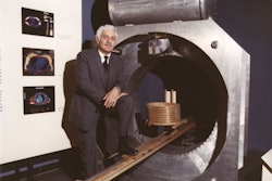
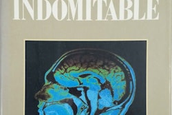
| Figure 2: A Machine Called Indomitable by Sonny Kleinfield, 1985. |
Raymond Damadian suggested in 1972 that NMR could be of value in medical diagnosis. He showed that different tissues emitted signals that varied in length, and that malignant tissues emitted signals that lasted longer than those from benign tissues. He filed a patent for this that was granted in 1974. By 1977 Damadian had completed construction of the first whole-body MRI scanner, described in the book A Machine Named Indomitable (figure 2).
In 1975 Peter Mansfield and Andrew Maudsley from Nottingham proposed a line technique, which in 1977 led to an in vivo cross section through a finger. In 1977 the Nottingham team succeeded in producing an image of a wrist. In April 1978 the Nottingham group produced high-definition images of a rabbit. They emphasized NMR would be able to provide both functional and anatomical information. By January 1979 they demonstrated recognizable anatomical detail in the living human forearm. They saw MRI as more than an alternative to CT scanning since it would become possible to achieve tissue discrimination and characterization through the analysis of the complex NMR signal.
 Figure 3: Early NMR of human head, 1980 (EMI). Image obtained with polar reconstruction similar to CT. The later images were improved by using the Cartesian (phase delay) reconstruction, as used today. Image courtesy of the archives of the British Institute of Radiology.
By 1978 Hugh Clow and Ian Young, working at EMI, reported the first transverse NMR image through the human head with rapid improvement of resolution (figure 3). More pioneering research was conducted at the University of Aberdeen under John Mallard. They developed the spin warp technique. Their team had published the first image through the body of a mouse in 1974. Peter Mansfield further developed the use of gradients in the magnetic field. He was a pioneer in the fast imaging techniques such as echo-planar imaging, which was introduced in the 1980s.
In the 1980s fast imaging techniques were also developed by Jurgen Hennig from the University of Freiburg, who introduced RARE (rapid acquisition and relaxation enhancement) imaging in 1986 (now better known as fast or turbo spin-echo imaging). Gradient-echo sequences were introduced by Axel Haase and colleagues from the Max Planck Institute in Gottingen. Roger Ordridge from Mansfield's group in Nottingham presented the first MRI movie in 1981. By June 1983 JM Kean and others from Nottingham showed what advances had been made by publishing MR images of the knee. Although the images appear primitive, there is excellent visualization of anatomical detail and a clear indication of what a powerful tool for musculoskeletal imaging MRI was to become. Figure 4 shows the MRI scanner at the Hammersmith Hospital from the early 1980s when I started there as a trainee registrar. There was a huge sense of excitement in the department.
 Figure 4: NMR scanner with cryogenic magnet at Hammersmith Hospital, dating from the early 1980s.
Dennis Carr from the Hammersmith Hospital MRI group and Wolfgang Schorner from Berlin published the first images in 1984 using the intravenous MRI contrast agent gadolinium DTPA dimeglumine in man.
References
Kleinfield S, A Machine Called Indomitable. Times Books, 1985.
Thomas A, Banerjee A, Busch U. Classic Papers in Modern Diagnostic Radiology. Springer Verlag, 2004.
Dr. Adrian Thomas is chairman of the International Society for the History of Radiology and honorary librarian at the British Institute of Radiology.
The comments and observations expressed herein do not necessarily reflect the opinions of AuntMinnieEurope.com, nor should they be construed as an endorsement or admonishment of any particular vendor, analyst, industry consultant, or consulting group.