With the help of FDG-PET, French researchers have discovered a common brain dysfunction among school-age children afflicted with fever-induced refractory epileptic encephalopathy (FIRES), according to a study published in the January edition of the Journal of Nuclear Medicine.
FDG-PET's ability to identify the pathological condition may pinpoint the origin and cause of FIRES, which previously has been unknown, according to lead author Michel Mazzuca, MD, from the neuropediatric department at Hospital Necker Enfants Malades in Paris (JNM, Vol. 52:1, pp. 40-47).
Study objective
Previous research has shown that brain abnormalities viewed on MRI are often limited to mesial-temporal structures and do not account for neuropsychologic findings. FDG-PET, however, has proved highly sensitive in detecting abnormal metabolic patterns in childhood epilepsy with negative MRI results.
The French study sought to detect hypometabolic areas in FIRES using a pediatric template and an age-matched population as a pseudocontrol group, and to determine the relevance of these areas relative to electroencephalograph (EEG) focus and neuropsychologic findings.
Eight patients, ranging in age from 6 to 13 years, were consecutively enrolled in the study. The mean age of first epileptic seizure was 5.8 years. All eight patients had undergone neuropsychological testing and FDG-PET imaging at the facility between 2003 and 2009.
The mean duration of subsequent epilepsy was 3.6 years. The researchers noted that at the time of PET imaging, no patient was seizure-free. The median seizure frequency was three per month -- with as many as 30 seizures per month -- with the seizures often occurring in clusters.
Imaging protocol
Brain MR images were obtained for all eight subjects using a 1.5-tesla scanner on the same day that PET imaging was performed with a whole-body scanner (ECAT Exact, Siemens Healthcare, Erlangen, Germany). PET images were acquired via 63 slices simultaneously, each with a 2.4-mm thickness in 3D acquisition mode. Patients also received FDG intravenously at a dose of 3.7 MBq/kg of weight.
The children were kept in the scanner for 30 minutes to acquire the PET images and were monitored for any head movements and adverse events, such as a seizure, stroke, or other negative reactions.
The study included a healthy control group, which mirrored the FIRES patients in age and had undergone the same FDG-PET procedure at the Paris hospital during the past seven years for focal epilepsy with negative MRI results. The 21 healthy subjects had a mean age of 11.4 years, ranging from 6.1 to 16.6 years, similar to the mean age of the patients in the FIRES group.
One pediatric radiologist, who had received information on the child's electroclinical data, analyzed the FDG-PET images independently.
Neuropsychologic results
The analysis showed that all FIRES subjects, with the exception of one patient, had reduced mental ability, with mental deterioration rated as severe in half of the eight patients.
The researchers found that neuropsychologic disorders primarily included language, frontal functions, behavior, and memory. Language disorders affected seven of the eight patients, while the remaining patient had no language at all. There were three cases of major receptive disorders, including one patient with auditory agnosia and three subjects with poor speech production.
 |
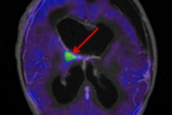
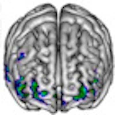
| Clinical images show spatial distribution of hypometabolic areas that are common to all patients in the FIRES group, illustrating bilateral network involving associative temporoparietal and orbito-latero-frontal cortices. The images compare eight patients in the FIRES group with the pediatric control group. Significantly, hypometabolic clusters are presented on the 3D mesh template in four orthogonal views (A-C and E) and on three orthogonal planes of standard brain (D). Image courtesy of the Journal of Nuclear Medicine. |
In addition, the authors noted that frontal disorders mainly consisted of lack of attention, slowness, and lack of response.
"Behavioral and emotional disorders of varying severity were associated in all cases," Mazzuca and colleagues wrote. "Memory deficit was evidenced in the three children with bilateral hippocampal atrophy who were able to perform the tests, [which] involved verbal as well as visual memory, immediate as well as delayed recall, resulting in marked episodic memory impairment."
Based on the MRI results, bilateral hippocampal abnormalities, including atrophy and hypersignal, were seen in three patients. One other patient had diffuse atrophy involving both neocortex and hippocampi. MRI, however, failed to disclose any abnormalities in the other four patients.
PET analysis
On visual PET analysis, all patients, including those with negative MRI results, disclosed several significant hypometabolic areas that were mostly severe and bilateral in seven of the patients and involved the neocortex (temporal in all patients, frontal in five patients, and parietal in four patients), as well as the temporomesial structures in seven of the patients.
"Surprisingly, no hypometabolism was detectable in temporomesial structures," Mazzuca and colleagues wrote. "In seven of eight cases, PET abnormalities were found in the posterior areas, mainly in temporal and parietal regions, but also in one patient spreading occipitally." In addition, EEG results were consistent with PET hypometabolic areas.
"PET findings do contribute to the identification of FIRES as a recognizable entity," the authors concluded. "Although the initial status epilepticus produces no evidence of neocortical necrosis, the functional impact is massive, and it persists for years."
By Wayne Forrest
AuntMinnie.com staff writer
January 3, 2011
Related Reading
Second PET scan can spot seizure origin in epilepsy patients, December 29, 2009
FDG-PET benefits findings in children with refractory epilepsy, July 2, 2009
3-tesla MRI both challenges and rewards pediatric radiologists, December 2, 2008
3-tesla MRI superior to 1.5-tesla MRI in epilepsy evaluation, October 1, 2008
Study shows benefits of very delayed cerebral FDG-PET imaging, December 21, 2007
Copyright © 2011 AuntMinnie.com