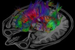
MRI exams can provide valuable information about neurological abnormalities in preterm infants. But the process of having an MRI exam can be hazardous to very fragile and unstable neonates, limiting the value of this powerful technology -- unless you have an MRI-compatible incubator.
MRI-compatible incubators preserve the environmental cocoon of the incubator, maintaining temperature and ventilation stability and reducing the need to handle infants in the MRI environment. But do these specialized incubators affect clinical management? Researchers at the Medical University of Vienna in Austria decided to find out; the results of their before-and-after comparative study were published in the September issue of the European Journal of Paediatric Neurology (2010, Vol. 14:5, pp. 410-417).
If you buy it, they will come
Purchase an MRI-compatible incubator and it will be used, the researchers found. MRI exams will be performed on more premature, low birth weight, and critically ill neonates, for whom the risks of the imaging exam may outweigh the benefits. And for the majority of infants, diagnoses will be more accurate and clinical management will probably change.
Because of the findings, neonatologists at the Medical University of Vienna now recommend that a routine MRI be performed for premature infants when metabolic disease or thrombosis is suspected, and in cases where injuries or malformations require surgical interventions.
The research team reviewed the medical records of unstable newborns in the hospital's neonatal intensive care unit (NICU) who had undergone an MRI exam during two 18-month periods: one period with and one period without the use of an MRI-compatible incubator. The NICU treated approximately the same number of patients during each time period. Information was analyzed regarding the number of exams performed, the time to perform each exam, the amount of sedatives needed, and any clinical management changes as a result of the MRI findings.
Thirty infants had an MRI procedure without use of an incubator between June 2003 and January 2005, of whom 17% weighed less than 2,000 g. Between June 2005 and January 2007, 99 infants had an MRI inside an MRI-compatible incubator (MR Diagnostics Incubator Nomag IC 1.5, Lammers Medical Technology, Luebeck, Germany), of whom 28% weighed less than 2,000 g.
The mean gestational age of patients decreased from 44 to 39.7 weeks with the use of the incubator. Additionally, incubator use increased from 14.8% to 36% for ventilated neonates.
How it's used
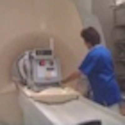
Infants are transferred from their incubator in the NICU to the MRI-compatible incubator, which has a built-in head coil that completely surrounds the infant's head. The incubator has auditory shielding, temperature and humidity regulators, a ventilation support system, and MRI-compatible monitors.
Lead study author Zsofia Rona, MD, reported that handling of the infant is reduced by half when an MRI-compatible incubator is used -- and the total procedure time is reduced by 20 minutes, on average -- because the step to stabilize the infant when placed on the MRI table is eliminated. During the 18-month time frame, this represented 33 hours of time in which the MRI suite could be used for other patients.
Additionally, the mean imaging time to scan the patient was reduced by four minutes with the use of the MRI-compatible incubator, even though an additional imaging sequence was performed when it was used.
Another advantage was that no procedures needed to be terminated due to insufficient sedation or infant instability. Without the incubator, 10% of the infants required additional sedation during the procedure.
Clinical management changes
The researchers combined both time periods being evaluated to analyze the effect of the MRI exams on everyday clinical practice. Management changes were initiated in 58% of the cases. The most common change in management involved the initiation or termination and choice of long-term anticonvulsive therapy or anticoagulation, operative decision-making, or antibiotic treatment.
The MRI exam provided additional diagnostic information in 43% of the cases, such as identifying underlying malformations or disruptive cerebellar development. For patients who had cranial ultrasounds performed after a clinical seizure, MRI identified a causative cranial pathology in 42% whose ultrasound findings were unspecific.
Following the MRI exam, the diagnosis was changed for all patients who had seizures of primarily unknown origins and for 86% of patients with central nervous system infections. The initial diagnosis was also changed in two-thirds of the patients who had cerebral malformations, and 73% of these patients had clinical management changes, as well.
The authors acknowledged the possibility of study bias being introduced due to increased neuroimaging of neonates in the second time period. There was a more than 300% increase in MRI procedures, as well as an increase in premature infants of lower gestational age and weight. However, the authors attributed this to the fact that an MRI-compatible incubator with an integrated head coil providing a better signal in the parietal regions of the brain can both provide a more accurate diagnosis and a safe environment for even the most unstable, critically ill premature infant.
By Cynthia E. Keen
AuntMinnie.com staff writer
August 31, 2010
Related Reading
Reduced brain tissue volume seen with postnatal dexamethasone, February 27, 2007
MRI predicts neurodevelopmental outcomes in prematures, August 17, 2006
MRI delineates brain injuries in newborns, May 3, 2006
MRI corresponds with neurodevelopmental outcome in preterms, November 14, 2005
MRI delineates complexity of preterm brain, April 30, 2004
Copyright © 2010 AuntMinnie.com