Although FDG-PET and MRI can contribute separately to the detection and assessment of carotid plaque, there is a lack of consensus between the two modalities when combined for hybrid imaging, according to U.K. researchers.
The researchers found that carotid plaque with high uptake on FDG-PET and a lipid-rich necrotic core on MRI was associated with increased metabolism and new vessel formation. However, they found no agreement on plaque instability between FDG-PET and MRI, which suggested separate pathogeneses or a different temporal evolution.
The challenge for physicians is to identify vulnerable plaque before it creates a blockage in the carotid artery that can lead to a stroke. Traditionally, MRI has been used to depict vulnerable plaque, while FDG-PET has been used to assess and quantify information within atherosclerotic plaques and, therefore, predict the probability of consequences from vulnerable plaque.
"We asked ourselves if there was a role for hybrid imaging ... and combined MRI and FDG-PET to tell us more about the plaque, improve characterization and plaque detection, and, thereby, provide insights into all that is going on inside these plaques," said lead author Dr. Leon Menezes from University College London. He presented research on the topic at the 2009 SNM annual meeting in Toronto.
Stroke consequences
Stroke is the third leading cause of death in the Western world and the leading cause of disability. It is caused by plaque in the carotid artery that becomes inflamed and ruptures to form a thrombosis. The thrombosis then moves along the artery and causes a blockage.
Thus, the goal of the study was to assess the feasibility of the multimodality hybrid imaging approach and explore the relationship between MRI's ability to identify the plaque, FDG-PET's capabilities, and the histological markers. The researchers used images acquired on separate scanners that were fused via software, rather than a combined PET/MRI scanner.
The researchers recruited 11 patients (10 men and one woman; mean age, 67 years) with symptomatic carotid disease. The patients received both an FDG-PET scan and MRI exam on the same day. One patient declined the MRI due to claustrophobia and could not contribute to the MRI data.
Imaging protocol
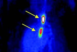
An FDG-PET scan of the neck was conducted in a single bed position, using 190 mBq of FDG and 90-minute circulation. "By drawing the region of interest, you can measure [standardized uptake value (SUV)] in the carotid artery and, by correcting for the background for measuring inside the internal jugular vein, you can determine the target-to-background ratio, which is one of the recommended means of assessing FDG uptake in atherosclerosis," Menezes said.
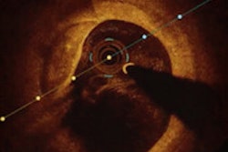
After the FDG-PET scan, patients were imaged on a 3-tesla MRI scanner with the aid of a head and neck surface coil and a time-of-flight angiogram to localize plaque.
Based on the study criteria, the definition of a carotid stenosis was blockage of at least 90%. The stenoses that were most "interesting," Menezes noted, were the ones that helped researchers describe the characteristics of the plaque. "Does it have a thin fibrous cap? A weak fibrous cap? And can you actually see the plaque becoming ruptured?" he said.
Among the 11 patients who underwent FDG-PET scans, the researchers found a statistically significant higher uptake in the symptomatic lesions compared to the asymptomatic lesions. "Moreover," he added, "FDG uptake was associated with microvessel density and increased metabolism."
Of the 10 patients who received an MRI exam, six were symptomatic with features of unstable plaque.
Modality dissociation
In comparing the two modalities, the reasearchers found "no association between FDG uptake and MRI signs of unstable plaque," Menezes said. "By virtue of the uptake, you could not distinguish which MRI lesions would be unstable."
From the data, the researchers concluded that carotid plaque with high uptake with PET is associated with increased metabolism and new vessel formation, while the MRI findings of lipid-rich plaques also are associated with increased metabolism and new vessel formation.
However, they found a dissociation in the results between FDG-PET images and the images obtained through MRI.
"There are some possible reasons for this, which we need to look into further," Menezes added. He suggested a separate process for each modality to look at factors related to the carotid plaque, "or there may be a different temporal evolution of a single process and each imaging tool is more sensitive to one phase or another."
Researchers from the University College London's Institute of Nuclear Medicine, Cambridge University Hospitals, Mount Vernon Hospital's Paul Strickland Scanner Centre, Brighton and Sussex University Hospitals NHS Trust, and Guy's and St. Thomas' NHS Foundation Trust participated in the study.
By Wayne Forrest
AuntMinnie.com staff writer
July 16, 2009
Related Reading
FDG-PET aids in detecting atherosclerosis activity, February 23, 2009
MRI beats PET for myocardial evaluation in patients with impaired LV function, January 9, 2009
FDG-PET/CT may play role in plaque evaluation, September 12, 2008
Cedars-Sinai explores PET's future in nuclear cardiology, April 11, 2008
Coronary CTA's time is now, according to Dowe, January 17, 2008
Copyright © 2009 AuntMinnie.com