Radiology has a deep pool of talent that spans the entire continent of Europe. This is one of the key conclusions to draw from the list of award winners for the 2022 EuroMinnies. The eight recipients of the individual and industry awards come from eight different European nations.
The winner of the Most Influential Radiology Researcher award comes from the same institution as the 2021 recipient, emphasizing the high quality of the research conducted at Erasmus MC in Rotterdam, the Netherlands. Also for the second year running, the Best New Radiology Device award went to a low-field MRI scanner, underlining the strong revival of interest in these systems.
In the EuroMinnies awards scheme, the full list of semifinalists was based on nominations submitted in late 2021 by members of AuntMinnieEurope.com. An expert panel comprising members of the editorial advisory board, past winners, and regular columnists selected two finalists in each category to go forward to the finals of the EuroMinnies. The judges then chose the winners in a second ballot.
During March, the winners will receive their trophies, and we will publish commemorative photos.
Most Influential Radiology Researcher
EuroMinnies 2022 winner: Dr. Gabriel Krestin, PhD, professor emeritus of radiology, Erasmus MC -- University Medical Centre Rotterdam, the Netherlands
For the second year running, this award has been won by a radiologist from Erasmus MC, after Dr. Marion Smits, PhD, was voted Most Influential Radiology Researcher in 2021.
"This underscores the high quality and impact of our research in Rotterdam -- something to which I have dedicated the past 25 years," said Krestin, who retired as head of radiology at the end of last year. "For both of us, this award reflects a group effort by all the talented and successful researchers in our department."
 Dr. Gabriel Krestin, PhD, from Rotterdam.
Krestin thinks a curiosity to find solutions to unsolved problems is the first prerequisite for a researcher.
"On top of that you need motivation, hard work, and endurance. Don't give up if it doesn't work at first try. And recognize that failure is part of the game -- you cannot always have positive results," he said. "More than anything, collaborate. Research only thrives in excellent groups; you cannot do it on your own. The environment counts -- an environment that supports research and a culture that values it."
Krestin admits that the days when he performed lab work, etc., lie far in the past. His main influence has been on building teams, deciding on the choice of topics, and supporting staff. He has also helped build the right infrastructures and mindset.
Krestin's own research was on MRI of the abdomen, but his department has succeeded in many fields, especially noninvasive cardiac imaging studies with new generations of CT scanners based on cooperation with cardiologists.
"Population imaging is another field that was more or less created here," he said. "Investing in equipment for the large-scale imaging of healthy elderly subjects and children and collaborating with epidemiologists became a highly successful field of research."
The multicenter MR CLEAN study introduced intra-arterial thrombectomy in stroke patients into routine practice and was an important game changer, according to Krestin. Also, molecular imaging and close collaboration with nuclear medicine has led to many opportunities, and researchers have developed multiple image processing algorithms, allowing automated assessment of biomarkers.
"The use of deep learning and artificial intelligence substantially reduces development times and improves the quality of algorithms," he noted. "We are moving into the field of data-driven precision medicine based on sophisticated predictive analytics for diagnosis, treatment selection, monitoring, and forecasting outcomes."
Another promising area is theranostics, i.e., finding biological substrates of disease and targeting them for diagnosis and treatment with radioisotopes. It starts with basic chemistry and radiobiology and requires collaboration with many disciplines, including pharmacy for creating new compounds, Krestin added.
He admires and respects Dr. Alexander Margulis and Dr. Sam Gambhir, PhD -- "two great personalities ... one is a really important historical figure and the other one died much too early," he said.
Krestin's advice to young researchers is to never give up.
"That doesn't mean you should do the same thing over and over again. Learn that success is not always granted and don't be discouraged if you don't succeed at the first try," Krestin said. "Look over the edge: the most exciting research topics can be found in areas between disciplines. Try to collaborate with clinicians, engineers, biologists, chemists, and computer scientists. Learn their language and respect their knowledge."
He plans to keep active in his retirement, particularly with mentoring.
"There is nothing more rewarding than seeing young researchers and clinicians strive and become successful and to know that you have contributed to that," he said, adding he will get more involved with startup companies. "I hope my experience can help young innovators to bring their ideas to market. I love to see how fast they move, how motivated they are, and how much they may contribute to healthcare."
Medicine and technology are converging, and radiology can drive innovation, Krestin predicts. "We have to stay open to integrating data from different sources like pathology, laboratory medicine, and all kinds of 'omics.' The shortage of personnel will further drive this transformation and offer unprecedented opportunities to future generations of researchers."
Runner-up: Dr. Denis Le Bihan, PhD, NeuroSpin, French Atomic Energy Commission (CEA), Paris
Most Effective Radiology Educator
EuroMinnies 2022 winner: Dr. Bettina Baessler, professor of clinical radiology, University Hospital Würzburg, Germany
The most important personal quality for an educator is a strong desire for lifelong learning and getting everybody to participate, Baessler told AuntMinnieEurope.com.
"Protecting your own knowledge is not the way to go. We should always be humble, patient, friendly, and open to new ideas," she said. "I really like the idea of a teacher as a 'guide on the side' instead of a 'sage on the stage.' The teacher should not take center stage."
 Dr. Bettina Baessler from Würzburg. Courtesy of Henner Huflage.
The first online course that had a long-term impact on Baessler's routine clinical work was a shoulder MRI course held by musculoskeletal radiologist Dr. Ravi Padmanabhan, director of Radiology Education Asia.
"It was completely different and included a step-by-step approach to reporting a shoulder MRI with hands-on training and real cases. He is kind and patient, and eager to let his students participate," she noted. "It very much inspired me in my own online course formats on the LernRad platform."
In the pandemic, Baessler has had to rapidly switch to online learning, building on her previous work on YouTube videos, the teaching program Cologne, and the Conrad platform of the German Roentgen Society (DRG).
"Although I like the online format, I really miss the experience of being with my students and personally interact with them," she explained. "Since my teaching style is very interactive, I cannot fully reproduce this in a purely online setting. We try hard, but full interaction is only possible in person. So, I am really looking forward to re-entering the auditorium in the near future."
Baessler entered radiology because she wanted a subspecialty that covered many different patients, clinical questions, and body parts, as well as having academic aspect. She loves the combination of modern techniques and digital applications and the broad nature of the field. Also, her cousin, an experienced radiologist in private practice, advised me to become a radiologist.
Above all, radiology education must be fun, and teaching helps to shape the field and attract talented young doctors to radiology, she continued.
"While I teach, I always learn something myself. It is getting the increasing recognition it deserves," she commented. "Teaching is not something you just have to fulfill because you are doing your residency at an academic institution, but it is a way to promote your specialty."
Having recently relocated from Zurich to Würzburg, she said it feels good to be back home, but she misses the skyline of Zurich and the Alps.
"A major difference between Switzerland and Germany is the amount of protected research time," Baessler said. "In Switzerland, it is quite standard to have at least some time for research. In Germany, this usually is only possible if you have secured your own funding. This makes it much harder to do excellent research, especially for novices."
The work culture is different too. In Zurich, for example, she often went for a coffee with colleagues. "This was really nice since it was quite easy to get in touch even with colleagues with whom you were not working together all day long. I think that most Germans would consider this "inefficient" (and maybe, it is), but I really liked this kind of social interaction during the workday," she said.
Baessler is convinced the future of radiology education is bright.
"We have always been a discipline a bit ahead of its time, and we are a leading discipline now when it comes to online education," she said. "If I didn't feel very positive about the future, I wouldn't have founded an online learning company in 2021 with my colleagues Mareike Franke and Nienke Hansen."
Runner-up: Dr. Francesco Sardanelli, University of Milan, Italy
Radiology Rising Star
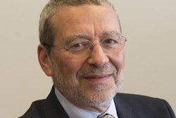
EuroMinnies 2022 winner: Dr. Paola Clauser, PhD, radiologist, Medical University of Vienna, Austria
During her 30s, Dr. Paola Clauser, PhD, made a career switch from Udine, Italy, to Vienna, and it's been a chastening experience.
"The transition was not easy -- sometimes it still isn't. It was not just about moving to a different country, but also from a small university in a small city to one of the biggest universities and hospitals in Europe in a capital city. I really had some hard times in the first few years," she said.
 Dr. Paola Clauser, PhD, from Austria.
To be able to report in German, she did many months of intensive courses, and she was helped greatly by her colleagues.
"The reporting style here is very structured and schematic, which allows me to learn fast how to write meaningful, understandable reports," Clauser explained. "The big challenge was to learn medical terminology and anatomy again. While Latin terms are accepted, German words are more common, and they rarely resemble Latin terms. Improving my German and my reports is still very much a work in progress."
She admits she entered radiology by chance. She wanted to do something that involved an interventional component. In her second year of residency at Udine, she started working in breast imaging, and she never looked back.
"All the consultants I worked with in Udine have had a strong impact on my career," she said. "The chief of the department at the time, Prof. Massimo Bazzocchi, was an inspiration for all of us. He taught me the importance of talking with the patients and listening to them."
In Vienna, working with her colleagues, in particular Prof. Dr. Pascal Baltzer, taught her that research is challenging but also exciting and fun.
Clauser co-founded the European Society of Breast Imaging (EUSOBI) Young Club (EYC) with Prof. Katja Pinker, but the idea came from Prof. Francesco Sardanelli.
"The aim is to give a voice and support young radiologists and to promote breast imaging," she pointed out. "We were enthusiastic about the project, and we were lucky to get a lot of help from many people on the way. They share with me the honor of this award -- as do my colleagues in Vienna."
The plan is to continue the group's successful series of webinars and online events at ECR and the EUSOBI annual meeting.
"We have a lot of ideas, so we will need more help, and there will be a call for new EYC committee members soon. I will have to leave the Young Club at 40. Till then, I hope I will be able to keep on working with them, either as a chair or as a committee member."
As for her own research, Clauser is finalizing the data analysis on a prospective study of fluoroethylcholine as an alternative tracer in PET-MRI of breast cancer patients. Part of the study has already been presented at the RSNA.
"I'd like to look more in-depth into the topic of tracers for advanced breast cancer staging. I am part of the EUSOBI diffusion-weighted imaging (DWI) working group, and with the pandemic seemingly giving us more freedom again, I plan to continue with multicentric collaborations on DWI standardization," she remarked.
Clauser is also focusing on the clinical role of contrast-enhanced mammography and gathering evidence for new guidelines. "What excites me the most, though, is screening and the move towards personalized screening. Early diagnosis is still one of the most powerful weapons we have against cancer."
She said she enjoys being directly responsible for patient management. Also, the work of a breast radiologist is very deeply connected with all the other specialists working in breast diagnostics and therapy, she added.
It's impossible yet to assess the impact of the pandemic on screening, but the team in Vienna is already seeing the effect on symptomatic patients, many of whom delayed visits and are now presenting with more advanced cancers.
According to Clauser, "Many organizational and communication aspects could have been managed much better during the pandemic. But one always knows better afterward."
Runner-up: Dr. Michail Klontzas, PhD, University General Hospital of Heraklion (PAGNI), Greece
Most Influential Radiographer
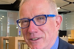
EuroMinnies 2022 winner: Jonathan McNulty, PhD, associate professor, University College Dublin School of Medicine, Ireland
When it comes to naming key issues in radiography, McNulty cites two: recognition and definition. It's only been four years since the European Commission's European Skills/Competencies, Qualifications and Occupations (ESCO) group formally acknowledged radiographers as healthcare professionals.
 Jonathan McNulty, PhD, from Dublin.
"The global recognition of our profession has really been a thorny issue," he told AuntMinnieEurope.com. "In fact, a number of countries in Europe continue to lobby for the establishment of university study that results in a degree. Partly the problem is the name: in some countries, the word 'technician' is used for radiographers, and any nomenclature with this term in it tends to be downgraded. Indeed, it is viewed by ESCO as being at a level below that of 'professionals'. Add to that fact that there are more than 50 titles for radiographers in Europe alone, and the situation can be quite confusing."
At the upcoming ECR meetings in March and July, McNulty will co-chair with European Society of Radiology (ESR) President Prof. Dr. Regina Beets-Tan a "Patients in Focus" session that will include talks from patients, radiologists, and radiographers regarding how the relationship between radiology professionals and patients can be enhanced. He will also facilitate a workshop of the European Federation of Radiographer Societies (EFRS) in partnership with Radiography focused on reader and reviewer outreach, as well as lead a session on the EFRS radiographer education, research, and practice project that will address the future of the field.
What are the growing edges in the field of radiography? There are a few, McNulty noted.
"Radiography is always exciting, as the technology is constantly evolving," he said. "There's an ongoing discussion about the role of artificial intelligence in radiography, as well as how to further establish more patient-centered protocols. And the issue of advanced radiography practice -- including reporting of findings and performing interventional procedures -- are also areas of widespread interest."
"In any case, we need to make sure that any advanced practice in radiography is underpinned by postgraduate-level education and training and that there are clear governance and quality assurance processes in place." he added.
It was the combination of technology and the opportunity for patient interaction that drew McNulty to a career in radiography back in the 1990s. At school, he had an interest in healthcare, but at the time wasn't yet clear what direction he would take. When he encountered the field of radiography at University College Dublin (UCD), he was hooked.
"I went to UCD, found myself studying radiography, and while I found it challenging, I loved it," he said. "It was a great mix of technology and patient care, with some great lecturers and clinical teachers, and I knew it was a career I could enjoy."
After university, McNulty found a full-time position as a clinical radiographer at Beaumont Hospital, a large teaching center, also in Dublin. During his time there, he specialized in interventional radiology and MRI, received a graduate diploma in MRI from Trinity College Dublin, and began to teach radiography part time at UCD; in 2005 he went full time. He earned a PhD in diagnostic imaging at UCD in 2014 and is currently vice principal for teaching and learning at the university's College of Health and Agricultural Sciences and Associate Dean in its School of Medicine.
McNulty's scope of influence further expanded in January, when he began tenure as the editor in chief of Radiography, replacing Prof. Julie Nightingale, PhD, of Sheffield Hallam University. The timing was fortuitous: He had recently served as president of the EFRS and his term on the board ended this past November, freeing him to take on the position in the new year. (McNulty previously contributed to the journal as a reviewer and as its associate editor.)
The editor-in-chief role brings McNulty deep satisfaction.
"One of the first things I remember when I started at UCD was picking up issues of Radiography and reading the articles," he said. "The journal informed my clinical practice as a student and a young professional and prompted me to start researching and writing myself."
McNulty plans to focus on supporting and expanding Radiography's reader engagement and impact in the field.
"My hope is that researchers who might initially think to publish on radiography topics in nonradiography journals, such as medical physics journals, -- because they are perceived to have more impact -- will choose to publish in Radiography instead."
In his free time, McNulty enjoys Ireland's national sports, Gaelic football, and hurling -- he is a supporter of the Dublin teams -- and coaches his son's Gaelic football and hurling teams as well. And although the COVID-19 pandemic has been distressing, it has had a silver lining: McNulty, his wife (who is also a radiographer), and their two children have taken to doing more walking in Dublin and its environs together.
"I love spending time with them, and we all really enjoy going for family walks," he said.
Runner-up: Charlotte Beardmore, Society and College of Radiographers (SoR), U.K.
Most Significant News Event in European Radiology
EuroMinnies 2022 winner: Dutch debate pros and cons of contrast-enhanced mammography and MRI for screening women with dense breast tissue
Should screening of dense breasts be done with MRI and not with contrast-enhanced mammography (CEM)? This question lay at the heart of a heated discussion that took place in the Netherlands throughout 2021 and still awaits resolution.
The debate has implications for breast imaging across Europe, and it looks set to come to prominence on 2 March 2022, when a special pros and cons session is devoted to contrast-enhanced breast screening at the ECR Overture online meeting.
 Dr. Ritse Mann, PhD. Photo courtesy of EUSOBI.
Dr. Ritse Mann, PhD, who is responsible for clinical breast research at the radiology department of Radboud UMC in Nijmegen and also who also works at the Netherlands Cancer Institute in Amsterdam, will speak in support of the motion: "This house believes that intermediate risk screening should be done with breast MRI and not with CEM.” Mann is an executive board member of the European Society of Breast Imaging (EUSOBI), a member of the scientific advisory board of the Dutch cancer society (KWF), and course director of the annual Nijmegen advanced breast imaging course.
In the opposite corner is Dr. Marc Lobbes, PhD, a breast radiologist at the Zuyderland Medical Center and the Maastricht University Medical Center in the Netherlands. During the 2 March session, he will argue that "this house believes that intermediate risk screening should NOT be done with breast MRI but with CEM."
The upcoming debate promises to be a lively and intriguing affair, but the matter is unlikely to be resolved until further evidence and data are available. According to Lobbes and Mann, a new trial is being designed by the former collaborators of the DENSE trial (November 2019, New England Journal of Medicine), which involved senior Dutch MR radiologists.
"At this point there is a call for such a study, and potential research groups are looking into that," Lobbes and Mann told AuntMinnieEurope.com on 16 February 2022 in a joint statement. "However, we are experiencing challenges in fulfilling the requirements for that call and are looking into the most optimal study design that gives answer to all the questions raised, but designing such a large study requires time to perform, and the COVID-19 pandemic did not help in that regard either. Right now, we don’t have any real outcomes to report, but further news is expected in late spring/early summer."
It is unclear which proposal is likely to be accepted and what the follow-up study will look like. These studies must be large and require a lot of resources, so plotting them takes time.
One thing's for sure: this debate looks certain to rumble on throughout this year. The outcome is eagerly awaited by the European breast imaging community. According to Mann, EUSOBI will soon provide new recommendations for screening of women with extremely dense breasts. Watch this space.
Runner-up: Ransomware attack on Irish health service causes disruption
Scientific Paper of the Year
EuroMinnies 2022 winner: Magnetic resonance imaging incidents are severely underreported: A finding in a multicentre interview survey. Kihlberg J et al, European Radiology, 20 July 2021.
When Johan Kihlberg, PhD, started work as a radiographer at the Center for Medical Image Science and Visualization (CMIV) in Linköping, Sweden, in 2004, he didn't expect MRI safety to be a prime area of focus for him. However, he and his colleagues are now playing a prominent role in the efforts to boost safety across Europe.
"I've been very lucky," he told AuntMinnieEurope.com. "I think this will catch an eye on MRI safety improvements. That's important, I think."
 On a mission to improve MRI safety. From the left: Johan Kihlberg, PhD; Annika Hall; Anders Tisell; and Peter Lundberg. The other author, Boel Hansson, is missing from this photo.
Kihlberg and his fellow authors found that MRI-related incidents tend to be "greatly" underreported, which would lead to catastrophic outcomes. Their results were published in July in the award-winning European Radiology paper.
The group found that 38% of incidents in the Swedish poll were reported formally, suggesting that about 100 incidents remained unreported. Radiofrequency burns were included, but attitudes reflected in the interviews suggest that the MRI personnel expect that burns in patients happen occasionally, leading to lack of reporting.
The authors also found that MRI knowledge increased because of the availability of MR physicists at clinical sites and that "almost all" radiographers were also concerned about MR safety knowledge for their respective radiologists.
While Kihlberg himself has not been a victim of a major incident involving MRI in his 22 years as a professional, he said it was a struggle to figure out best safety practice at first.
"At the time, I found that there were some features that you had to tweak a little bit," he said. "You can set the scanner in certain modes so that you can scan patients in ways you couldn't before. Fifteen years ago, you couldn't scan patients who had a pacemaker, but now we scan these patients every week. The whole context has changed, and we need to follow the changes."
Since the paper's submission, Kihlberg and colleagues have started a group consisting of radiographers, radiologists, and physicians who come together to develop guidelines and give educational lectures on MRI safety. These talks align with the paper's call for educational efforts needed to enhance a culture of safety. While the lectures have been held in Sweden only, Kihlberg said they have garnered interest from radiology departments in Norway and Denmark.
The team is now planning a follow-up study that will focus on MRI safety measures outcomes among a larger cohort.
"It [safety] is a shared responsibility. We have, of course, the radiographer being the closest person, and we have physicists who can give us support," he commented. "But the most responsible person is the radiologist. That's the person who should be doing the risk-benefit analysis. Everybody, though, has some responsibility at different levels."
Runner-up: Impact of specificity on cost-effectiveness of screening women at high risk of breast cancer with magnetic resonance imaging, mammography and ultrasound. Kaiser CG et al, European Journal of Radiology, 29 January 2021.
Best New Radiology Device
EuroMinnies 2022 winner: Swoop portable MRI scanner, Hyperfine
The winner of this year's EuroMinnies award exemplifies the revival of a debate between low-field and high-field MRI that many observers thought had long ago been settled in favor of the latter.
The Hyperfine scanner is a 0.064-tesla scanner in an era where the upper boundary of field strength has hit 3 tesla, with even 7-tesla systems being debated for clinical use. With its system, Hyperfine has gone the other direction, developing Swoop as a device that's so small and portable that it can be wheeled directly to the patient's bedside.
 The mobility of the Hyperfine Swoop is a key benefit. Image courtesy of Hyperfine.
The permanent-magnet system is 1.45 m tall and weighs just 630 kg. Due to the small size of the scanner, Swoop is intended primarily for brain imaging, but it also can be used for imaging extremities such as the wrist, knee, and foot.
Hyperfine sees its technology as having the potential to revolutionize radiology by making MRI more accessible in locations where it never could have been used previously. Indeed, one of the company's founders is Jonathan Rothberg, PhD, who also founded Butterfly IQ, a developer of handheld ultrasound scanners.
In addition to sending Swoop scanners to the patient's bedside, the portable systems could be installed in ambulances as part of a mobile stroke unit, similar to how small CT scanners are being used today. The company has also received grants to study the feasibility of installing its systems in developing countries that lack access to advanced imaging like MRI.
The vendor also sees the scanner being used in place of imaging modalities that deliver ionizing radiation, like CT, as part of an effort by physicians to reduce a patient's lifetime exposure to radiation.
Hyperfine received U.S. regulatory clearance for Swoop in August 2020, and it is in the process of working with notified bodies in Europe to apply for the CE Mark. Scanners have been installed at several locations in the U.K. and Europe, and the company has begun building a European sales organization, first targeting the U.K. as well as Pakistan.
Runner-up: Somatom X.ceed CT scanner, Siemens Healthineers
Best New Radiology Software
EuroMinnies 2022 winner: QP-Prostate AI software for prostate MRI, Quibim
QP-Prostate is an artificial intelligence (AI) application developed by Spain's Quibim to help clinicians during prostate MRI reporting -- from visualization to quantification. Ultimately, the software is meant to help clinicians save time diagnosing prostate cancer, the second most common cancer in men.
An estimated 9.6 million people worldwide died from prostate cancer in 2018, and the incidence of the disease is increasing in most countries, according to the World Health Organization.
"Prostate MRI images are some of the most difficult exams to read, and some tasks of the prostate read are laborious, such as contouring and measuring the prostate," said Angel Alberich, PhD, Quibim's founder and chief executive officer. "Reporting on a prostate exam can take somewhere between 30 and 45 minutes. QP-Prostate can do this in seconds."
 Angel Alberich-Bayarri, PhD, and some of the Quibim team outside the company's offices in Valencia, Spain.
QP-Prostate enables automated segmentation of the transitional zone, peripheral zone, and seminal vesicle regions of the prostate, which leaves more time for clinicians for image interpretation and diagnoses, Alberich said. The software received 510(k) clearance from the U.S. Food and Drug Administration (FDA) in March 2021, with clearance pending in Europe.
The software runs as a client-server model that requires a high-performance computer installed by Quibim inside the hospital or medical clinic network. This server communicates with PACS networks through DICOM protocols, and QP-Prostate is thus accessible through the web browsers of any standard "off-the-shelf" computer connected to the network.
The software can communicate with any PACS via standard Query/Retrieve DICOM operations. Once the complete prostate MR study has been received, QP-Prostate checks the available sequences.
"Radiology is all about pattern recognition. The more experience, the better you get. QP-Prostate is doing exactly this. It has been trained by a vast quantity of data and therefore is excellent at recognizing patterns. It decreases the intervariability we see between radiologists and helps them focus on detecting abnormalities," Alberich said.
In order for QP-Prostate to be used to analyze an MRI exam, the study must include at least the following:
- T2-weighted MR sequences and diffusion-weighted imaging (DWI) or
- T2-weighted MR sequences and dynamic contrast enhanced (DCE) or
- T2-weighted MR sequences, the DWI, and the DCE
QP-Prostate automatically runs processing steps such as automatic arterial input function (AIF) selection, registration and motion correction, and spatial smoothing algorithms to improve image quality and minimize heterogeneity across vendors.
The software includes a DICOM viewer to improve the review process of a single study and its related image sequences. In addition, the viewer has basic image manipulation and segmentation capabilities and advanced segmentation ones for prostate. Lastly, Alberich noted, the software includes a structured reporting option for filling a standard PI-RADS template (Prostate Imaging Reporting and Data System version 2.1).
With the aim of receiving clearance for QP-Prostate in Europe, Alberich and colleagues are currently working with several selected partners to further optimize and validate the software.
"We are putting QP-Prostate through a rigorous validation process with multiple centers," he said.
Looking ahead, Alberich said Quibim is working to position itself as the "go-to" imaging partner for diagnostics and drug discovery. Future products will include products based on real-world evidence with applications across oncology/immunology, rheumatology, and neurology.
"You will see Quibim bringing new radiology AI software in these areas while we grow our positioning in the space of clinical biomarkers and companion diagnostics," he commented.
Runner-up: Medical Open Network for AI (MONAI), Nvidia and King's College London
Best New Radiology Vendor
EuroMinnies 2022 winner: Radiobotics, Copenhagen, Denmark
Unlike many artificial intelligence (AI) software developers that have focused on more sexy applications such as detecting cancer or stroke on advanced imaging modalities, this Danish firm has chosen to focus on musculoskeletal imaging and the oldest imaging modality: radiography.
It's shaping up to be a fortuitous decision.
 The team at Radiobotics celebrate the news of their award with a special cake.
"A lot of hospitals and clinicians have now been working with CT and MRI (AI) technology, and they're ready to try other modalities and look for other pathologies," Chief Operating Officer (COO) Stine Mølgaard told AuntMinnieEurope.com.
Radiobotics was incorporated in September 2017 by co-founder CEO Mads Jarner Brevadt, PhD, with three other co-founders – Chief Technology Officer Pavel Lisouski, Chief Scientific Officer Martin Axelsen, and COO Stine Mølgaard -- all officially joining the company by early 2018.
The company developed the prototype for its first AI algorithm -- RBknee -- in collaboration with the radiology department at Bispebjerg and Frederiksberg University Hospital in Copenhagen. Suitable for orthopedic or radiology settings, RBknee analyzes a standing posteroanterior knee radiograph to identify common radiographic findings associated with the diagnosis of osteoarthritis (OA): osteophytes, subchondral sclerosis, and joint space narrowing. Its machine-learning algorithms then conclude whether OA is present.
RBknee received the CE Mark in early 2021, and a different version of the software received U.S. Food and Drug Administration (FDA) 510(k) clearance in August 2021.
Radiobotics has benefited from a variety of partnerships with both academic research institutions and a variety of other companies. In addition to its relationship with Bispebjerg and Frederiksberg University Hospital, Radiobotics has partnered with U.S. teleradiology services provider Vrad since 2020 for training data and software validation. In December, that relationship was extended to include development of algorithms to enhance the speed and accuracy of bone fracture x-ray studies.
Also in December, Radiobotics signed an agreement to make RBknee available on image exchange software provider TeleRay's platform. In June 2021, Radiobotics was selected for GE Healthcare's Edison Accelerator program, which is aimed at supporting early-stage companies developing healthcare AI applications.
Other Radiobotics partners include: Charité University Hospital in Germany; the East Midlands Imaging Network (EMRAD) in the U.K. National Health Service (NHS); Oxford University Hospitals in the U.K. NHS; Erasmus University Medical Center in the Netherlands; and the University of Texas Medical Branch in the U.S.
The firm is now working on other musculoskeletal radiography AI algorithms in joint analysis, trauma, and quality-control applications. For example, RBmarker is designed as a quality-control tool for x-ray laterality markers, while RBfracture evaluates x-rays for hip fractures.
"The vision for Radiobotics is to fully automate everything in the image," Mølgaard said.
The company has submitted RBfracture for the CE Mark, and it hopes to launch the application this quarter. RBfracture is the first in a total of 11 new applications expected to be introduced by the third quarter as part of one platform.
"In order to solve substantial problems, you also need to have a substantial product," she said.
Runner-up: Lucida Medical, Cambridge, U.K.