In forensic cases, multislice CT (MSCT) now plays a crucial role in comparative identification, reconstructive identification, and lesion identification, and has important advantages over dry bone analysis, according to French researchers.
One of MSCT's major assets in forensic anthropology is the elimination of lengthy bone preparation, which may damage fragile bone, and this can be particularly useful when bones are very burned or charred, noted Dr. Fabrice Dedouit, PhD, from the Service de Médecine Légale, Hôpital de Rangueil in Toulouse, France, and colleagues. Also, the modality allows virtual access to bones where physical access is impossible, such as bones embedded in sediment or concrete blocks, and of major interest is conservation of the images and the reconstructions.
 Postmortem multislice CT explorations of two different unknown bodies, with reconstructions in volume-rendering technique mode permitting detection of metal. Above: Chest level: Detection of metallic foreign bodies secondary to surgery of the neck (metallic surgical clips due to surgery of the thyroid) and the chest (metallic wire on the sternum secondary to heart surgery). Below: Left foot: Detection of two surgical screws within the first metatarsal secondary to surgery. Images courtesy of Dr. Fabrice Dedouit, PhD.
"Further studies can be carried out on the object scanned, independently of its state of preservation. This opens up a new approach to quality control and expert supervision, as well as image transmission and use in forensic telemedicine," they wrote in an article posted online on 14 November by the British Journal of Radiology (BJR).
MSCT can be performed in the country where the bones or body were discovered, while further work on image analysis and reconstructions can be continued in another country. Image and data processing offer objective visualization and recapitulation of forensic results, with the high spatial resolution of MSCT. The main drawbacks to routine use of MSCT in anthropology, however, are the limited accessibility of such systems, their cost, and the real need for the radiologist to be familiar with anthropological techniques, the authors wrote.
For MSCT to produce optimal results, many technical conditions must be adhered to in terms of image quality, spatial resolution, and contrast; if these are not met, the final images may be unusable, particularly for analysis of fine trabecular bone lesions. The initial CT must be performed with appropriate voltage, amperage, field-of-view, and slice thickness, and after acquisition, reconstruction time is critical, they explained.
Choice of thickness and interval and choice of filters influence voxel size and the possibility of radiological interpretation, and postprocessing requires powerful computers. Choice of reconstruction technique may include 2D reconstructions (multiplanar reconstructions: sagittal, coronal, oblique) or 3D reconstructions (maximum intensity projection, surface shaded display, or volume rendering technique), and must be appropriate.
"The wide range of possibilities offered by MSCT may seem bewildering to the nonradiologist!" Dedouit et al stated. "Concerns relating to imaging of the bones and teeth of deceased persons are not very different from the concerns of clinical practice: The radiologist must always be aware of the reasons behind the examinations in order to use an appropriate CT image acquisition protocol."
When a body is not identified or is unidentifiable -- as with skeletal, charred, putrefied, or mutilated individuals -- radiological investigation is necessary, and its major aims are identification of the person and of the injuries. This can be important in "hidden crimes" such as a person killed by gunshot and then burnt in their home. Comparison of antemortem radiographs or MSCT images with postmortem MSCT images can be vital for positive identification of the deceased: This is the goal of comparative identification, they explained.
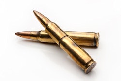
In ballistic trauma, MSCT can help to determine criteria such as location of the projectile(s) and determination of the direction of fire plus entry and exit bone wounds. Ricochets may also be detected by imaging, and the same is true of sharp trauma, with visualization of deep bone laceration or of cartilage section in some cases.
"MSCT is effective in identifying infectious lesions, and is more informative than plain x-rays. Infectious processes can affect bones, teeth, and also soft tissues, so MSCT can be very helpful in study of mummies," the researchers noted. "More rarely, bone tumors may be observed, or features secondary to metabolic changes or anemia such as cribra orbitalia, Harris lines, osteopenia, or osteoporosis. Foreign bodies may also be detected within bones."
Another advantage of MSCT is detection of the stigmata of occupational stress, or cultural artifacts such as intentional or artificial cranial deformations. Other bone lesions may be diagnosed such as arthropathies that may be important for comparative identification.
Taphonomy and pseudopathology must not be forgotten, they stressed. Forensic taphonomy is the interdisciplinary study and interpretation of postmortem processes of human remains in their depositional context, and taphonomic details are important for estimating time since death and differentiating injuries from postmortem changes. Radiologists who perform postmortem MSCT must be familiar with late postmortem changes, to avoid misinterpreting normal changes as traumatic injuries, and such confusion, especially when exhumed bodies are concerned, may have significant judicial consequences.
Many anatomical parts may be involved: postmortem changes of the ossicular chain of the middle ear, axial and appendicular joint disarticulations. Due to loss of soft tissues and costal cartilages, the ribs, sternum and clavicles may collapse into the chest cavity. The hyoid bone may fall near the spine, while the mandible may be disarticulated and some teeth may be absent because they have fallen after death. This must not be confused with post-traumatic lesions, according to Dedouit and colleagues.
They advocate a multidisciplinary approach in such work because it involves communication and data exchange between radiologists, forensic pathologists, anthropologists, and radiographers.
Also posted online by BJR on 14 November was an article about advances in postmortem CT angiography, the lead author of which was Dr. Silke Grabherr, PhD, from the University Center of Legal Medicine Lausanne-Geneva, University Hospital of Lausanne in Switzerland. Look out for a follow-up report next week on AuntMinnieEurope.com about this second BJR article .
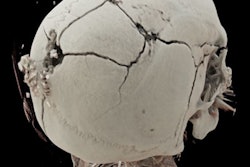
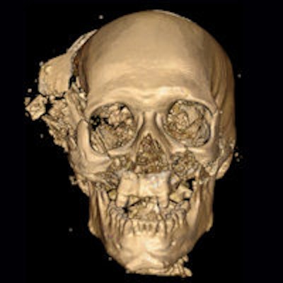
Editor's Note: The image on the home page is a volume-rendered 3D CT reconstruction conducted after a homicidal death. The image shows the characteristic bony and metallic fragments on the exit side of the skull, where the bullet caused a large loss of brain, leading to shattering of the skull. Image courtesy of Dr. Giuseppe Guglielmi.