High-quality CT images of the abdomen and pelvis can be acquired by using the latest iterative reconstruction techniques at an ultralow dose (1.3 ± 0.8 mSv) in patients with Crohn's disease, enabling the detection of most complications, according to Irish researchers.
Patients with Crohn's disease are at risk of significant cumulative effective doses of ionizing radiation from imaging, primarily CT, and protracted exposure to low-level ionizing radiation is a real concern because there is an existing lifetime risk of developing small-bowel lymphoma and other intestinal malignancies, noted Dr. Siobhan O'Neill, from the department of radiology at the University College Cork in the Republic of Ireland. This is compounded in patients with more severe disease by the use of immunomodulatory drugs, and the potential carcinogenic effects of significant cumulative radiation exposures is a serious concern.
"Patients with Crohn's disease often require diagnostic imaging, be it for initial diagnosis, monitoring of therapeutic response, or perioperative evaluation," she stated. "These patients can be subject to serial imaging over prolonged periods of follow-up due to early age of presentation and, sometimes, decades of active disease."
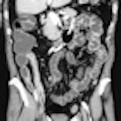
 Small-bowel CT enteroclysis. Crohn's disease extended throughout the ileum, with thickened wall and typical enhancement of the mucosal layer. Fibrofatty proliferation of the mesentery, also known as creeping fat, is visible. Image courtesy of Dr. Gian Rollandi of Ospedali Galliera in Genoa, Italy.
CT has a high sensitivity and specificity for detecting luminal and extraluminal disease and complications, and yields images with high temporal and spatial resolution with a short acquisition time compared with conventional enterography and MRI. However, a standard CT of the abdomen and pelvis (CTAP) exposes the patient to a radiation dose of about 8 mSv (literature range 3.5-25 mSv). The radiation exposure from CTAP is substantial compared with chest radiography (0.02 mSv), plain film of the abdomen (0.7 mSv), and barium studies (3.5-5 mSv), O'Neill explained in a presentation at the RSNA 2011 meeting in Chicago.
Disease-specific low-dose CT protocols can help limit radiation exposure, but the inherent contrast resolution required to resolve soft tissues that have only slightly varying attenuation values can have a negative impact by increased image noise, which can affect image quality. The reconstruction algorithm used can have an impact on image quality, she added. CT images are traditionally reconstructed using filtered back projection (FBP), a rapid and efficient method with relatively low mathematical demands, but FBP is not well-suited to low-dose protocols.
Iterative reconstruction, a noise-efficient reconstruction algorithm, minimizes the impact of increased image noise in images acquired at low doses, improving image acceptability. This technique allows radiation dose reduction by reducing image noise compared with the standard FBP technique. When it is used in low-dose CT scanning in patient and phantom studies, a radiation dose reduction of 32% to 65% is possible with significantly less noise and preserved low-contrast resolution and image quality, according to O'Neill.
"The next step in optimizing image quality is the development of advanced generations of iterative reconstruction, such as model-based iterative reconstruction. This can incorporate a physical model of the CT system into the reconstruction process to characterize the data acquisition process, including noise, beam hardening, and scatter. It offers the potential to further enhance image quality at even lower radiation doses," she noted.
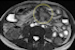
Typical indications for CT in Crohn's disease include establishing the initial diagnosis and quantification of disease distribution, extent, and activity, as well as assessment for complications such as active enterocolitis, abscess, fistula, stricture, and lymphadenopathy.
In an article published last year, O'Neill recommended limiting the use of those imaging studies that involve ionizing radiation to clinical situations in which they are likely to change management. She also advocated implementing advances in low-dose CT technology, and, where feasible, using alternative imaging modalities, such as ultrasound or MRI (Clinics and Research in Hepatology and Gastroenterology, February 2011, Vol. 35:2, pp. 105-110).
CT remains the cornerstone technique in studying small-bowel diseases, and provides a means for panoramic examination of the organ in the context of other parts of the gastrointestinal tract, which is important because pathologies elsewhere can mimic the symptoms of small-bowel diseases, according to Dr. Gian Rollandi, a radiologist from Ospedali Galliera in Genoa, Italy. CT images have high spatial resolution and are suitable for revealing subtle pathological changes, such as small tumors and vascular abnormalities. They are also reliable for identifying truly negative cases, which is very important in the clinic, he noted.