CT scanning has had inestimable effects on medical care, and we owe a huge debt to Godfrey Hounsfield and Allan Cormack for their pioneering work. It is difficult to overestimate the impact that the EMI/CT scanner has had on medicine.
The neuroradiologist James Bull said in 1977 that Hounsfield's invention was the most important advance in radiology since Wilhelm Röntgen radiographed his wife Bertha's hand in November 1895. Within five years of the introduction of CT, there were machines throughout the world, and the technique was rapidly adopted into clinical practice. It is now difficult to imagine how medicine was practiced before the introduction of CT scanning. Shown below is the brochure for the EMI 1010 scanner, "the most advanced diagnostic system for neuroradiological examinations" (Figure 1).
 Figure 1. Brochure cover for the EMI 1010 scanner, signed by Godfrey Hounsfield. All images courtesy of Dr. Adrian Thomas.
The history of the development of CT is complicated. The need for tomography lies in the fact that in conventional radiography a 3D object is displayed as a 2D image with no depth information.
Classical tomography was well developed by the 1950s, the pioneer work having been done by Bernard Ziedses des Plantes and published in the 1930s. In classical tomography, the x-ray source and the detector both move, and the points of pivot all lie in the same plane. Objects out of the plane are blurred and those at the level of the pivot are in focus. This tomographic image gives information in one plane and helps in diagnosis.
The award of the Nobel Prize for Medicine and Physiology to Godfrey Hounsfield and Alan Cormack in 1979 emphasized the arrival of the new technique of CT; however, there were many others who had worked in this field. The mathematical basis for image reconstruction started with Johann Radon. In 1917, he published his work on the "Radon transform" with the idea of reconstructing a function from a set of projections, therefore playing a significant role in the development of CT.
In the mid-1940s, Shinji Takahashi in Japan worked on the principles underlying rotational radiography and developed sinograms. In 1958, Boris Korenblyum and his co-workers built a medical CT scanner in Kiev. In 1960, William Oldendorf developed experiments to demonstrate the feasibility of CT scanning using a rotating phantom made of nails and mounted on a track. In the early 1960s, David Kuhl and Roy Edwards developed an apparatus for emission CT scanning. It is very interesting to observe the number of disconnected workers considering the same problem but coming from different directions.
Allan Cormack developed the basis of what became CT scanning, as well as a mathematical approach for looking at the problems of variations in body tissues that are important in radiotherapy. Cormack considered a fine beam of radiation passing through a body. In 1957, he conducted experiments using a phantom that had circular symmetry.
By 1963, Cormack was ready to try experiments on a phantom that did not have circular symmetry. The apparatus consisted of two cylinders containing a detector and a gamma ray source. The phantom lay between the two cylinders, and the work was completed in the summer of 1963. Cormack then considered how many measurements needed to be made, because only a finite number of measurements can be made with beams of a finite width. The results were presented for publication in graphical form and were published in 1964. There was almost no response to the publications, and certainly there was no commercial interest.
In the 1960s, Godfrey Hounsfield was working at EMI Ltd. in Hayes, Middlesex, U.K., and was interested in pattern recognition. He considered a closed box with an unknown number of items inside that could be looked at from multiple directions using a radiation source and a radiation detector. The results of the transmission readings could then be analyzed by a computer and presented as a slice in a single plane.
Hounsfield developed a mathematical approach in a process of reconstruction. The original apparatus resembled that used by Cormack and was a simple lathe holding the object to be examined. The early experiments used Perspex phantoms of varying complexity. Shown below is an image from an album by Godfrey Hounsfield (Figure 2). Scanned directly from the original Polaroid photograph, it was labeled by Hounsfield and is the first ever CT image taken by him. It took nine days to take the picture and 15 minutes of computing time to reconstruct.
Following the use of Perspex phantoms, a section of human brain in a Perspex box was examined. Most of the pictures from the lathe bed were scanned in 1969 and 1970. The original lathe bed apparatus may be viewed in the library of the British Institute of Radiology in Portland Place in London.
 Figure 2. Polaroid image of the first CT scan made by Godfrey Hounsfield of a Perspex phantom.
Hounsfield looked at practical applications and approached the Department of Health in London in 1968. He met Dr. Cliff Gregory and Gordon Higson, who were scientific advisers at the Department of Health and Social Security (DHSS). Hounsfield was then introduced to Dr. Evan Lennon, a radiological adviser who knew Frank Doyle from the Hammersmith Hospital. Doyle gave Hounsfield two lumbar vertebrae of different densities, which Hounsfield examined and returned to Doyle with computer printouts of numbers in the coronal plane of the vertebral body. Hounsfield had already worked out a scale of numbers, and Doyle was impressed with the result.
Lennon also made contact with two other radiologists: Dr. James Ambrose and Dr. Louis Kreel. Ambrose was a neuroradiologist from Atkinson Morley's Hospital in South London, and Kreel was from the Royal Free Hospital; Kreel subsequently moved to Northwick Park Hospital in Harrow, where he did pioneer work using the EMI body scanner. Ambrose later discovered that Hounsfield had been previously dismissed by an eminent radiologist as a crank.
It became apparent that EMI would not spend any more money without the support and a contribution from DHSS. Work continued on specimen radiography, and then on 14 January 1970, a meeting was held at DHSS with the three radiologists, as well as Lennon, Gregory, and Higson. The initial results were promising, and they agreed to produce a prototype brain machine to be located at Atkinson Morley's Hospital.
The prototype scanner was installed at Atkinson Morley's Hospital on 1 October 1971. It is quite remarkable that Hounsfield went in one jump from the primitive lathe bed apparatus to the prototype CT scanner.
The prototype looks very similar to our modern CT scanners, and the original is on display in the Science Museum in South Kensington, London. The scanning time was four minutes per slice, and the slice thickness was a little more than 1 cm. There was no computer attached to the machine, and the data were taken on magnetic tape by car to be analyzed by EMI at Hayes. The data were reconstructed using an ICL 1905 mainframe computer and a picture with an 80 x 80 matrix that took 20 minutes to reconstruct. The data could have been reconstructed using a 160 x 160 matrix; however, this would have taken much more time.
The first patient scanned was a 41-year-old woman with a possible frontal lobe tumor. The data were acquired and the tapes were sent to EMI, with results returned after two days. The cystic tumor in the left frontal lobe was clearly shown (Figure 3), and Ambrose said the result caused Hounsfield and himself to jump up and down like football players who had just scored a winning goal.
The scan is presented in the opposite direction from modern scans and is viewed as a neurosurgeon would look, which is from above. Modern scans are viewed as looking from below upward. The reverse of the Polaroid has the words "original 1st patient scanned." The preliminary results were presented at the 32nd annual congress of the British Institute of Radiology, which was held at Imperial College in April 1972. The paper produced a sensation, and the first press announcement was in The Times on 21 April 1972.
EMI then started the production of a brain machine and made five: one for the National Hospital, Queen Square; one for Manchester; one for Glasgow; and two for the U.S. (one for the Mayo Clinic and the other for Massachusetts General Hospital). All machines were installed in the summer of 1973. Radiology was changed forever.
 |
| Figure 3. Polaroid image of the first clinical CT scan. |
CT scanning has continued to develop since the 1970s. Spiral CT represents a significant advance in the technology of CT scanning and has significantly increased its clinical value. The first clinical cases and performance measurements were presented as a work-in-progress by Willi Kalender, Peter Vock, and Wolfgang Seissler at the 75th anniversary meeting of RSNA in 1989.
The development of spiral CT and multislice scanning was made possible by the advances in computing. Data no longer have to be taken away for analysis but can be reconstructed almost instantaneously. There are amazing results with modern multislice scanners with improved spatial resolution, with virtual endoscopy, and faster scanning enabling complex dynamic studies. This dramatic improvement was made possible because of the provision of higher continuous x-ray power and improvements in computers.
References
Thomas AMK, Banerjee AK, Busch U. Classic Papers in Modern Diagnostic Radiology. Berlin, NY: Springer Verlag; 2005.
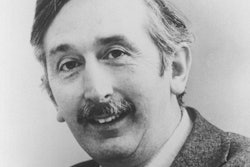
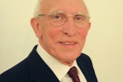
Dr. Adrian Thomas is chairman of the International Society for the History of Radiology and honorary librarian at the British Institute of Radiology.
The comments and observations expressed herein do not necessarily reflect the opinions of AuntMinnieEurope.com.