A multidisciplinary team from the Technical University of Munich (TUM) has successfully tested a nano-CT device on a velvet worm and created 3D x-ray images at resolutions of up to 100 nanometers (nm). The device promises much for virtual histological applications, but work must continue on small animal models and human samples, according to the team.
The TUM Nano-CT system is based on a newly developed x-ray source, which generates a particularly focused beam, without relying on x-ray optics. In combination with an extremely low-noise detector, the device produces images that approach the same resolution as a scanning electron microscope while also capturing structures beneath the surface of the object under investigation, the authors stated in their paper published in the 21 November edition of Proceedings of the National Academy of Sciences of the United States of America (PNAS).
 Euperipatoides rowelli, a species of velvet worm. Copyright: de Sena Oliveira/Universität Kassel/reproduced with permission from PNAS.
During a CT analysis, the object under investigation is x-rayed and a detector measures the respective amount of radiation absorbed from various angles. Three-dimensional images of the inside of the object can be constructed based on several such measurements. However, high-resolution capture of very small objects has been hampered due to the current limitations of the technology.
Devices with x-ray lenses can achieve high resolutions, down to 50 nm and even smaller at synchotron facilities, but the size and type of material of the sample is limited when using x-ray optics. Meanwhile, with a standard micro-CT, without x-ray optics, the resolutions so far have been limited to 0.5 to 1 micron (µm). The TUM device can achieve higher resolutions than these, according to the authors. This means it can image smaller samples than a standard device with good resolution, but is also more adaptable to different sample sizes and materials.
"Our system has decisive advantages compared with CT, which uses x-ray optics," said Mark Müller, doctoral candidate at the chair of biomedical physics at TUM and lead author of the PNAS article. "We can make tomographies of significantly larger samples and we are more flexible in terms of the materials that can be investigated."
Speaking to AuntMinnieEurope.com, he explained how the technology not only provides insight into the evolutionary biology of arthropods, but also holds important implications for medicine, pointing to its use for examining healthy and tumorous tissue samples. Work on tumor tissue is ongoing and the results have yet to be published, he added.
"With the help of the nano-CT we can perform virtual histology," Müller said. "While in conventional histology thin slices are examined in 2D with a microscope, we can image relatively large tissue samples and retrieve similar or additional information about the tissue in three dimensions. This can improve pathological diagnosis, and the sample preparation and data acquisition is less complicated and time-consuming than the standard histological procedure."
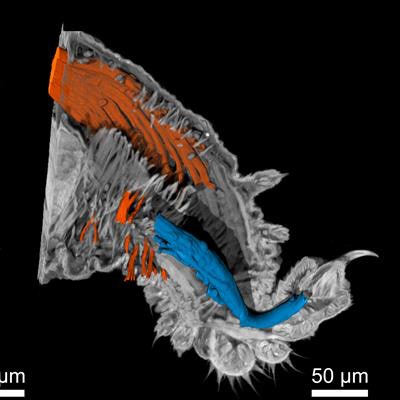
 Nano-CT images of a velvet worm leg. Left: Surface of the leg. Right: A view inside the tissue with muscle fibers highlighted. Copyright: Müller, Pfeiffer/TUM/reproduced with permission from PNAS.
The group is already working with small animal models and human samples with the sample size for a single measurement typically not much larger than a cube with a 2 mm edge length.
Because the nano-CT prototype remains a long way from commercialization, it may be too speculative to talk about cost benefits. Currently, histology studies are performed on histological slices that are individually stained and examined. This is a long and costly process; therefore the availability of a nano-CT machine in clinics should make histological processes more efficient, according to Müller.
"Nano-CT offers the possibility to provide 3D high-resolution data from biological and biomedical samples. In clinical diagnostics, the nano-CT data can provide new insights into the microscopic development of widespread illnesses such as cancer and facilitate the work of histopathologists," he noted.