Visibility of colon polyps is increased and sensitivity improved with the use of threshold-based color-coding software in virtual colonoscopy (also known as CT colonography or CTC), according to a new study from Italy. Even flat lesions were more often visible with use of the open-source 2D-based interpretation software.
Although low-dose CTC protocols with fecal tagging are effective for polyp detection, some smaller lesions -- and particularly flat polyps -- have been difficult to diagnose at CTC. Detection is even more challenging using mAs settings due to high image noise and the small CT density difference between the lesions and the colonic wall, said Dr. Lorenzo Faggioni from the University of Pisa in Italy.
To address this lack of conspicuity, some medical image processing applications, such as open-source OsiriX software (OsiriX, Geneva), generate color lookup tables (CLUT) that can enhance the contrast between lumen and wall of the colon, revealing CT density differences by means of color gradients (color coding), Faggioni explained in a presentation at the 2010 European Congress of Radiology (ECR).
Using the software's inverse thresholding function, CT values corresponding to colorectal polyps typically appear pink, tagging materials as violet, and the colonic mucosa a dark green.
The researchers aimed to "assess the feasibility and usefulness of color coding for primary 2D reading of CTC images with OsiriX," Faggioni said.
They examined the results of 30 previously interpreted virtual colonoscopy exams (17 men, 13 women; mean age, 63 years; range, 44-81 years) presenting with 52 proven colonic lesions, including:
- 10 flat lesions
- 4 polyps < 6 mm
- 20 polyps 6-10 mm
- 13 polyps 10-30 mm
- 5 colonic masses
Patients underwent a same-day fluid tagging regimen using iodinated oral contrast. Low-dose VC was performed prone and supine using a 64-detector-row CT scanner (LightSpeed VCT, GE Healthcare, Chalfont St. Giles, U.K.) set at 120 kV, 50 mA, rotation time of 0.5 sec, 1.375:1 pitch, 0.625-mm collimation, and a reconstruction interval of 1 mm using standard and soft reconstruction kernels.
Datasets were exported in DICOM format to a Macintosh computer running OsiriX version 3.5.2. Two experienced readers working in consensus evaluated the data with primary 2D interpretation of axial images using:
- A standard bone visualization window (300 HU level, 1,500 HU width) and no CLUT
- Then, a customized visualization window (-220 HU level, 1,800 HU width) using a version of CLUT known as GEcolor
Lesion conspicuity scores with and without color coding were compared by means of a two-tailed Wilcoxon signed-rank test. Lesion conspicuity was expressed through a semiquantitative scale from 1 (poor) to 4 (excellent). The correlation between maximum lesion size and differences in lesion conspicuity with and without color coding was assessed using a Spearman rank test.
"Lesion conspicuity was significantly higher with the color coding rather than without it, and we saw these results over and over," Faggioni said. "The conspicuity of all lesions was higher with the color coding than without it. Even in a subgroup of flat lesions we saw that visibility with color coding was much better than without color coding."
|
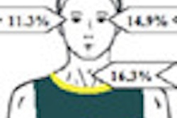
Analysis using Wilcoxon score ranking shows higher overall conspicuity with the use of CLUT color coding.
Lesion conspicuity was rated better in 20/52 lesions (38.5%), equal in 30/52 lesions (57.7%), and worse in 2/52 lesions (3.8%), Faggioni said. The researchers found a significant inverse correlation between lesion conspicuity improvement with color coding and lesion size (rs [relative strength] = -0.4219, p = 0.00184).
 |
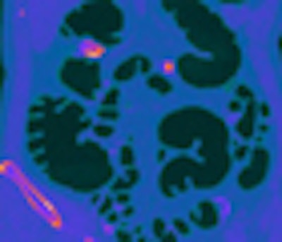
| A pedunculated polyp is better visualized using OsiriX-based color coding. All images courtesy of Dr. Lorenzo Faggioni and Dr. Emanuele Neri. |
"The use of color coding for primary 2D reading of CTC images can improve visualization of colonic lesions, especially of small polyps and flat lesions, which are harder to see with standard CTC," Faggioni said. "OsiriX should be considered a promising alternative to commercial image processing software solutions for color coding-assisted analysis of CTC datasets with color coding."
 |
| A flat lesion in the ascending/transverse colon is detected only using the threshold-based color-coding software technique. |
Responding to a question from the audience, Faggioni said that the nonlinear inverse transfer function was the most reliable way to use the software for polyp color coding, because it "enhances the contrast differences between the various voxels, especially between the wall voxels and the voxels of structures outside the wall," he said. "We also tried to use the linear threshold, but this was not as effective."
By Eric Barnes
AuntMinnie.com staff writer
April 6, 2010
Related Reading
CAD catches most flat polyps on virtual colonoscopy, December 16, 2009
Sophisticated electronic bowel cleansing boosts polyp detection, August 3, 2009
Topographical height mapping moves mountains in polyp detection, July 27, 2009
Image tools cut false positives in unprepped VC, August 4, 2008
OsiriX founders look to take software to next level, March 25, 2008
Copyright © 2010 AuntMinnie.com