BARCELONA - Coronary artery disease is a clear and present danger in the U.S., affecting an estimated 16 million Americans and killing about 500,000 of them each year. In the U.K., coronary artery disease is responsible for one in four deaths.
The situation is brighter in Spain, where Mediterranean diets and lifestyles may contribute to what has historically been one of the lowest rates of heart disease in the Western world. But the popularity of fast food and smoking may signal a gloomier future. A study a few years back, for example, found unexpectedly high levels of atherosclerotic plaque in young adults from Barcelona.
The need to evaluate growing populations at risk for coronary artery disease is only expected to increase the demand for coronary CT angiography (CTA), which has of late become the go-to modality, favored for its speed, accuracy, reproducibility, and noninvasiveness compared to gold-standard conventional angiography.
But coronary CTA isn't as easy to perform as it could be, according to Chunliang Wang from the Center for Medical Image Science and Visualization at Linköping University in Linköping, Sweden. At a presentation at the 2008 Computer Assisted Radiology and Surgery (CARS) meeting this week, Wang discussed the problem of hidden coronary arteries and introduced a novel segmentation method that might make them easier to examine.
Using thin-section 64-slice CT, "the exam produces hundreds of slices, so if you want to give a diagnosis you have to go through all of them," he said. "We need an easier reconstruction method that can give you more information and is less time-consuming."
A handful of 3D techniques are available to help physicians make a decision faster -- for example, maximum intensity projections (MIPs) and volume rendered (VR) reconstructions, which have proven their worth in CTA of the carotid and femoral arteries, for example. But they don't work as well in the heart, Wang said.
"Getting contrast to the coronary arteries also enhances the ventricle, and these two structures are overlaid on each other, so it's difficult to give a diagnosis on 3D," he said. And because the enhanced structures have similar intensity, traditional segmentation methods such as thresholding yield unsatisfactory results.
"We want to remove the ventricle and you can do this manually, but we are trying to develop an easier method," he said. The software was developed in cooperation with researchers at Uppsala University in Uppsala, Sweden.
 |
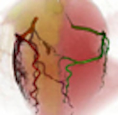
| In coronary CTA, many artery segments are hidden by the heart chambers, and the similar CT densities of these structures make it difficult to segment the arteries automatically. All images courtesy of Chunliang Wang. |
To visualize a solution to the reading problem, imagine two flowers growing in a single flowerpot, Wang said. Their roots will grow into each other like arteries and veins in the heart. The origins of each root are hard to tell apart. But if two plants are pulled apart, the roots can be separated according to their connectivity to one plant or the other. Similarly, he explained, his group's segmentation method interactively segments and virtually "pulls apart" the arteries for easier viewing while retaining their original spatial associations.
The software was built as a plug-in for OsiriX, an advanced open-source PACS workstation (OsiriX Foundation, Geneva, Switzerland), Wang said. The segmentation function is based on an optimized virtual contrast injection algorithm that uses the "fuzzy connectedness" of the vessel lumen to separate the contrast-filled structures from each other.
In contrast to conventional algorithms, the fuzzy connectedness approach doesn't define precise vessel borders. That approach is unsatisfactory because "there's always a risk you might make a mistake on the results," he said. "In our algorithm we try to include some low-intensity soft tissue surrounding the vessel, and we just focus on separating the objects, so you can be more confident about your diagnosis."
The seed-growing approach facilitates preservation of detailed information about the coronary artery tree, Wang said. If you try to define precise vessel borders, you can never be sure if a stenosis is real or merely a result of the processing, he said. In their study, every vessel retained at least one pixel of soft tissue surrounding it.
The researchers evaluated the algorithm in 42 clinical coronary CTA datasets acquired with isotropic voxels (0.3 mm to 0.5 mm) on a 64-detector-row CT scanner.
 |
| Above, a fuzzy connectedness theory is introduced to segment different components by the connectedness relationship. In this theory, suppose p is a path joining a and b, the lowest grayscale value of pixels on this path is assigned to be the strength of path p. In an image there are many possible paths between a and b. But the method uses the strongest path to represent the relationship between a and b, and the strength of this particular path is assigned to be the connectivity value between a and b. In order to use the fuzzy connectedness theory for segmentation (below), the user first supplies two sets of seeds. Then, for each pixel in the image, the user can decide its membership by comparing the connectedness strength from the point to the different seeds. For example, if v is more tightly connected to seed 1, then v is said to belong to object 1; otherwise, it should belong to object 2. |
 |
 |
| The fuzzy connectedness theory can be used to segment different components in images in a process known as skeletonization. If a path p joins a pair of points, a and b, every point on the path will contribute a strength to hold its neighbors together, and the strength of the whole path is determined by its weakest point. As a result, if the strength used to drag the two points a and b passes over the weakest point, the path will be broken. In 3D images there are millions of possible paths between a and b; this method assigns the connectedness strength between a and b as the strongest possible path. |
"After the segmentation, each pixel is actually pointing to its strongest neighbor in the tree structure," Wang said. "We try to repeat the stretching for every structure." Using the graphic user interface, seeds are "planted" and "grown" for each vessel in the tree, he said. The cases were processed in a median of 6.4 minutes.
According to the results, 100% of the main branches (right coronary artery, left circumflex artery, and left anterior descending artery) and 86.9% (219/252) of the evaluable minor branches were intact. Correct centerlines were obtained automatically in 94.7% (321/339) of the intact vessel branches.
 |
| Segmentation results (above) show the percentage of intact vessels following segmentation. The chart below shows the number of seconds required to process each step (time range in parenthesis). |
 |
 |
| Completely segmented coronary artery tree is seen in 3D image (top), which can be rotated to facilitate viewing. Tagged volume-rendered technique (VRT) image is below. In the volume-rendering interface, the colors and the opacity of the various objects can be changed. |
 |
The software postprocessing method is a promising tool for coronary CTA, providing good visualization of the coronary artery tree with minimal user interaction that will be reduced in further development, Wang said. "In the future we are aiming for a fully automated procedure, where you push a button and in a few minutes you open up the results," he said.
With further development, the process could be made more time-efficient than current thin-slab techniques, he added.
By Eric Barnes
AuntMinnie.com staff writer
June 27, 2008
Related Reading
3D development outpaces facilities' readiness, June 23, 2008
Mayo pares PACS limitations with intelligent image routing, June 11, 2008
Video game technology speeds beating-heart surgery, June 9, 2008
Software unifies control of multiple informatics applications, June 2, 2008
Using informatics to meet communication challenges, February 7, 2008
Copyright © 2008 AuntMinnie.com