Radiologists from a small city in southern Spain that is famous for its olive oil and medieval fortresses have collected two prestigious awards for their groundbreaking research in the emerging field of diffusion tensor imaging (DTI).
Judges in the poster hall at the RSNA 2018 congress in Chicago gave two magna cum laude awards to Dr. Teodoro Martín Noguerol and colleagues from the MRI unit at Clínica Las Nieves, Sercosa, Health Time in Jaén. Only 29 of these accolades were given to authors of the 2,758 exhibits shown at the meeting.
"These awards are just the tip of the iceberg for all the good work that is being developed in Spanish radiology," he told AuntMinnieEurope.com. "A large amount of public and private hospitals are committed to research and development, not only for conventional radiology but also for new advanced techniques that are allowing improved patient healthcare in common clinical practice."
The team collected the awards for its handbook of skeletal muscle DTI for beginners and a detailed description of how to optimize DTI acquisition for spinal cord assessment.
The aim of the handbook was to review the physical basis of DTI and its technical adjustments for skeletal muscle evaluation. The group explained, from an educational point of view, the biological meaning of parameters derived from DTI studies in the musculoskeletal system and showed potential applications of DTI for skeletal muscle evaluation in different clinical scenarios.
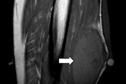
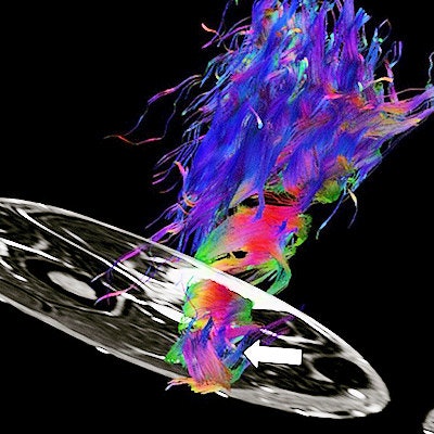
 A 50-year-old male with acute pain at the posterior aspect of the thigh during a football match. MRI shows severe semitendinosus tear (grade III) at morphological study (arrow). DTI reveals a severe decrease of fractional anisotropy (FA) values at injury site (arrow). Note the twist-shape appearance of hamstrings on DTI reconstruction with retraction of muscle fibers (arrows). All images courtesy of Dr. Teodoro Martín Noguerol.
Becoming familiar with the physical basis and technical adjustments of DTI for skeletal muscle assessment is essential for a proper sequence implementation and data interpretation.
Parameters derived from DTI studies provide valuable quantitative information about muscle fiber physiopathology.
"DTI reconstructions allow us to obtain a 3D representation of muscle fibers and can be used as a complementary tool for skeletal muscle qualitative assessment," Martín Noguerol and colleagues noted. "Nowadays, there is not a clear indication of how or when DTI studies should be performed for skeletal muscle evaluation."
DTI is showing promising results for muscle lesion characterization and also treatment monitoring, but further studies are needed to validate this technique in common clinical practice, they added.
Spinal cord evaluation and other applications
In their second poster, the investigators reviewed the physical basis of DTI and their technical adjustment for spinal cord evaluation. They discussed the biological meaning of parameters derived from DTI studies in the spinal cord and showed the potential applications of DTI for spinal cord evaluation in various clinical settings.
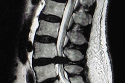
 A 65-year-old female with cauda equina syndrome. A cystic lesion is identified at the lower spinal cord. The marked decrease of fractional anisotropy values within the cystic component of ependymoma (arrow) leads to an absence of representation of fibers/white-matter tracts at this level. Note the thinning and displacement of the rest of tracts around the lesion. DTI has the added value for surgical planning and assessing the behavior of spinal cord tracts, which are displaced by the lesion.
DTI provides both valuable qualitative and quantitative information for spinal cord assessment that can be used as a complementary tool for conventional MRI spinal cord examinations, and it can help to confirm, rule out, and even detect pathology in patients with apparent normal morphological studies and persistence of clinical symptoms. However, the integration of DTI as another sequence in routine protocols for spinal cord assessment is unclear, and specific technical adjustments must be considered prior to acquiring DTI studies for spinal cord assessment.
A judicious use of DTI, adjusting field-of-view and acquisitions times in specific subtypes or myelopathies targeted lesions would be a good start point for introducing this promising sequence in clinical and radiological practice, the authors wrote.
Health Time has a long tradition in displaying educational exhibits and scientific posters at RSNA. It has received more than 30 awards in the last few years, including nine prizes at RSNA 2018. The group is headed by Dr. Antonio Luna, PhD, chief of MRI.
 Dr. Teodoro Martín Noguerol from Jaén, Spain.
"Being part of an international congress such as RSNA constitutes in itself an award," noted Martín Noguerol, a 37-year-old radiologist who has worked in Jaén since 2010. "I'm glad about the recognition that these awards entail, but the most important goal of these presentations is to share our knowledge and experience with the radiological community and open the chance to receive feedback from people who are interested in our work."
The researchers are working on clinical applications of DTI outside the brain because, although this technique has been used successfully for central nervous system evaluation, it has scarcely been explored in other parts of the body, he continued. In the case of the spinal cord, several studies have previously evaluated its feasibility for assessing trauma or degenerative related myelopathy, but tips and tricks on obtaining an adequate acquisition are of importance for both technicians and radiologists.
Another area of untapped potential is the application of DTI for assessing primary and secondary myopathies, especially those related to sports injuries.
"There is a promising horizon in the field of muscle injuries evaluation using quantitative data derived from DTI studies," Martín Noguerol pointed out. "We are now working on scientific papers that support the technical adjustments for DTI acquisitions proposed in these e-posters, especially in the field of spinal cord lesions and sport-related muscle injuries."