VIENNA - For high-quality trauma care, radiologists must recognize the importance of making the CT scanner a safe place for severely injured patients by working with the emergency department and anesthetic staff, making sure that protocols for scanning and reporting trauma patients are adhered to, and developing services that can be delivered within a few minutes 24 hours a day.
That's the central message of Dr. David Kessel, immediate past president of the British Society of Interventional Radiology and consultant radiologist at Leeds Teaching Hospitals, U.K.
Trauma patients can present anywhere. Even severe cases may need to be brought to smaller centers as a lifesaving staging post, so radiologists need to ensure that services are in place to perform and interpret imaging in these critically ill patients. Trainee radiologists must learn about trauma imaging by exposure both in the workplace and in teaching sessions.
 An injured 14-year-old boy is transferred by the resuscitation team in the x-ray department. Image courtesy of Dr. David Kessel.
"In trauma circles, the CT scanner used to be known as 'the doughnut of death.' The reason for this was a combination of logistics and technology," he noted. "The logistical impediment relates to needing to have a scanner appropriately located in relation to the emergency department and ready to receive severely injured patients within a very short time from their arrival in the hospital. This means that there must be radiographers available, transfers must be rehearsed, and the teams responsible for ongoing resuscitation are familiar with the CT environment and ready to accompany the patient during the scan."
The technological factor that has turned the CT unit into 'the doughnut of life' is the speed of scanning. A whole-body trauma CT can be performed in less than two minutes, and it gives information about all major injuries to the head, spine, thorax, abdomen, and pelvis, explained Kessel, a speaker at this afternoon's refresher course.
From the head to midthigh, CT should be the primary investigation of choice for the severely injured patient and should not be delayed. Experienced radiologists are a crucial element of an early and accurate diagnosis, he stated. Trauma scans should be reported according to simple algorithms, and the goal is to provide a primary survey report that identifies immediately life-threatening injuries within minutes of a scan. Next must come a more in-depth secondary survey assessing other important diagnoses that will have an impact on patient management.
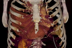
 External markings and abdominal distension due to the trauma. Image courtesy of Dr. David Kessel.
"Unfortunately, there is a lingering sentiment in many emergency departments that CT is a dangerous place for the critically ill patient. In fact, the CT department is as safe as anywhere else if it is properly equipped and the patient is adequately supported by the resuscitation team. There is no reason that ventilation, resuscitation, and monitoring should not continue during scanning," Kessel pointed out.
Diagnostic radiologists who are likely to see trauma patients need to consider how they will be able to provide this crucial service in a timely fashion. Realistically, this means considering how scanning and reporting will be performed within an hour of the patient arriving in the hospital.
However, Kessel admits some issues still await resolution. When scanning, the most important thing to remember is that there must be sufficient body coverage for the scan and intravenous contrast must be given. "When reporting, the most sensible approach is to ask everyone to step back whilst you are reviewing the scans to allow you to take stock and review the images in a logical fashion. Mistakes are most likely to be made if the radiologist is rushed or distracted by clinicians each with their own specific concern," he added.
The simplest way to prevent pitfalls is to scan and report trauma patients according to protocol and using standard pro formas, Kessel maintains. Leeds has a single protocol for trauma scans detailing the scan settings, contrast dose, and image reformats so that all scans are performed to a fixed recipe. In addition, there are two standard report sheets: one for the initial assessment, and another for the radiological secondary survey. Each sheet provides details about the body areas that need to be commented on.
Many trauma patients are young, and because trauma CT scans impart a significant radiation dose, there is a possibility of cancer-related deaths. This must be balanced against the risks to the patient from the trauma, he noted. There are two keys to minimize radiation dose: Ensure that patients are only scanned if they have a genuine indication with appropriate mechanism of trauma or constellation of injuries, and use the correct protocol and avoid repeat scanning.
"There is increasing evidence that early CT scanning improves outcome in trauma patients," Kessel stated. "The military experience in Camp Bastion in Afghanistan has led to a model in which incoming wounded military and civilians can be taken directly into the CT scanners on arrival at the military hospital."
He is also watching closely for the results of an important ongoing trial in the Netherlands that is randomizing trauma patients either to conventional therapy or CT before resuscitation. If this turns out to confer advantages by directing treatment to the most life-threatening aspects of a patient's injuries, there will be pressure to change the early trauma pathway. This will have a major impact for planning CT services, including their location and ensuring that they are staffed 24 hours a day, he predicts.
 Dr. Lars Lönn, PhD, is a professor of endovascular surgery and a consultant in the department of radiology at the National University Hospital of Denmark in Copenhagen.
The recognition and treatment of injuries have evolved dramatically over the past two decades, according to Dr. Lars Lonn, PhD, professor of endovascular surgery and consultant in the department of radiology at the National University Hospital of Denmark in Copenhagen. Image quality and acquisition times have improved, and imaging's use as a diagnostic tool is essential. It also serves as the bridge to endovascular minimal invasive procedures.
"Vascular and trauma surgery, as well as interventional radiology, are now truly in a transitional phase. Standard open repair is being replaced by endovascular and minimally invasive techniques to an extent far beyond our imagination," he commented. "The techniques developed for elective cases are also applicable for injured patients. The endovascular treatment for emergencies has consequences for both institutional and regional organization."
Lönn thinks success can only be achieved by having a team with wide elective experience, and most important, there must be good team spirit, where everybody remembers why they are working in hospitals: to improve a patient's healthcare.
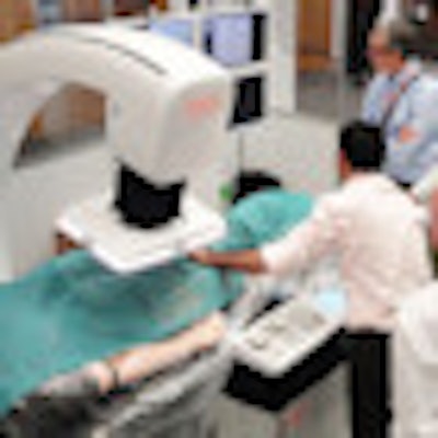
 Simulators can play an important role in facilitating training of embolization procedures, as shown by this photo from the Orzone Educational Sessions at the CIRSE 2011 meeting, held last September in Munich, Germany. The training setup consisted of the Orzone hybrid training cath lab; Orcamp, the Mentice endovascular simulator; VIST-C and the Laerdal full-body patient; and SimMan 3G. Image courtesy of Orzone AB, Göteborg, Sweden, and Dr. Lars Lönn, PhD.
To avoid problems, a safe chain of logistics, in which all team members collaborate and share knowledge, is essential. A change of mind-set and a more systematic approach are necessary to prevent errors, and he advocates the implementation of training in a simulated environment as an integrated part of daily practice. A combination of expertise in simulation technology and knowledge of trauma medicine/surgery will help to create a positive environment and safe medical care for emergency medicine. Thus, integrating appropriate training methods into daily practice will be crucial in the future.
"More collaboration between specialties will be a must," Lönn stressed. "It is a challenge to keep up with this future, and prognostication is a difficult moving target. In my 25-year career, the development of imaging modalities, more recently functional techniques, was not a consideration when I entered radiology."
The trauma surgeon and the radiologist need to embrace their collaboration in order to improve patient safety and satisfaction. Management of trauma patients should involve interventional radiology, neurosurgery, orthopedic surgery, plastic surgery, vascular surgery, anesthesiology, among others. Use of e-learning and validated curricula can strengthen team training, leading to better care in real-life scenarios, he concluded.
Originally published in ECR Today March 1, 2012.
Copyright © 2012 European Society of Radiology