Certain quantitative PET parameters can help clinicians predict how well patients with Hodgkin's lymphoma will react to their treatment, according to preliminary results from a study conducted in the Ukraine and presented at ECR 2018 in Vienna.
In a comparison of several variables, the researchers found that metabolic tumor volume and mean standard uptake values (SUVmean) were statistically significant indicators of event-free survival.
"The end-of-treatment PET quantitative parameters may be utilized to help detect patients at high risk of early Hodgkin's lymphoma relapse," said study co-author Dr. Mykola Novikov from the department of diagnostic radiology at the Israeli Oncologic Hospital (LISOD) in the Kiev region.
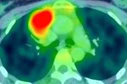
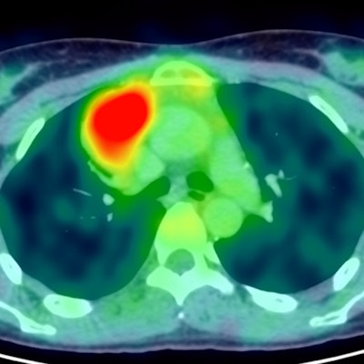
 Maximum intensity projection image of F-18 FDG PET/CT scan of a patient with Hodgkin lymphoma demonstrating hypermetabolic disease limited to the mediastinum. All images courtesy of Dr. Mykola Novikov.
Modality of choice
The use FDG-PET/CT is a well-established method for evaluating patients with Hodgkin's lymphoma and is considered the gold standard for imaging the progression of the disease. The hybrid modality also is the primary choice for staging, restaging, and response to therapy.
"Qualitative and visual assessment is considered to be the standard in multiple trials and in tailoring therapy, for example being incorporated in additional clinical decision-making," Novikov told ECR attendees. "But the quantitative approach remains a point of ongoing research and investigation regarding metabolic information in these types of patients."
This multicenter study was the first attempt by Ukrainian researchers to collect PET data on patients with Hodgkins' lymphoma from a variety of imaging centers.
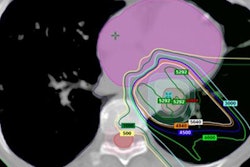
 Axial PET and fused PET/CT image of the same patient with Volume of Interest marking for quantitative assessment of F-18 FDG uptake in mediastinal adenopathy, providing values of different quantitative metrics.
"Unfortunately, we have to admit there is huge variability and inconsistency of imaging, which dramatically affects the eligible patient population, so we have produced only preliminary results for now," Novikov added.
The aim of this study was to retrospectively calculate and analyze metabolic quantitative PET parameters in patients with Hodgkin's lymphoma, and to evaluate possible correlations between quantitative PET indicators and event-free survival among these patients.
The researchers gathered 45 patients with Hodgkin's lymphoma; 53% of the cases involved either stage I or stage II disease, while the remaining 47% presented with stage III or stage IV disease.
As part of the treatment regimen, the patients received either doxorubicin, bleomycin, vinblastine, and dacarbazine (ABVD) treatment or a combination of bleomycin, etoposide, doxorubicin, cyclophosphamide, vincristine, procarbazine, and prednisone (eBEACOPP), which is typically used in Europe.
In addition, three PET scans were performed. The first exam was conducted at baseline; the second scan after two cycles of therapy; and the third exam at the end-of-treatment at least four weeks after therapy completion.
To determine the efficacy of quantification parameters, researchers used a fixed threshold segmentation method with a maximum standard uptake value (SUVmax) of 41%, as recommended by the European Association of Nuclear Medicine (EANM). From that point, the researchers calculated SUVmax, peak SUV, mean SUV, metabolic tumor volume, and total lesion glycolysis (TLG) at all three PET scan time points.
PET parameters
In reviewing the results, Novikov and colleagues found the overall response rate to cancer treatment was 95.5%, with 73% of patients with negative results and the remaining 27% with positive PET images on the second interim PET scans. At the end of treatment, negative predictive value changed to 82% and positive predictive value to 18% (p < 0.05)
A further analysis of PET parameters discovered which variables were most effective in predicting event-free survival. For example, a multivariate analysis revealed that positive predictive value was the only statistically significant factor in determining event-free survival with a hazard ratio of 1.07 (1.0 to 1.15, p = 0.00008).
Based on the Cox regression model, metabolic tumor volume and SUVmean based on the third PET scan were most statistically significant in event-free survival. Metabolic tumor volume recorded a hazard ratios of 1.07 (1.0 to 1.13, p = 0.005, while SUVmean registered a hazard ratio of 2.0 (1.4 to 2.82, p = 0.0001).
Conversely, quantitative metrics based on baseline and the second interim PET scans were not statistically significant in terms of determining the clinical outcome of the Hodgkin's lymphoma patients.
"In conclusion, the end-of-treatment PET quantitative parameters may be utilized to help detect patients at high risk of early heart gives lymphoma relapse," Novikov added.
The researchers cited several limitations of the study, which included the relatively small patient cohort and heterogeneous study population. They recommended additional investigation with more patients and follow-up PET scans at different intervals during cancer treatment.
"Also, we should try different quantification methods for comparison to find the most robust and valuable method to predict the most valuable quantitative indices," Novikov concluded.