Early results from an Italian-led clinical trial showing a multimodal tomographic system could improve breast cancer diagnosis were presented at ECR 2023.
Dr. Elena Venturini from San Raffaele Hospital in Milan discussed initial findings from the SOLUS project (Smart OpticaL and UltraSound diagnostics of breast cancer), showing that the system has high sensitivity and moderate specificity.
"The results confirm the possibility of acquiring useful information for the characterization of breast lesions," Venturini said in a session on 2 March.
While breast ultrasound is the go-to supplemental imaging method for suspected breast lesions found on screening mammography, Venturini noted that its specificity still needs to be improved in order to reduce false-positive rates.
Optical imaging is one idea that has been explored in this setting. When performed at multiple wavelengths, this method can gather information about tissue composition and blood parameters while also reducing scattering. This leads to acquiring information on microscopic tissue structures. However, Venturini said that diffuse optical imaging has limited spatial resolution, meaning morphological data from other techniques are used to build anatomical landmarks.
"Ultrasound could be the ideal method to derive anatomical information and complement directly observed therapy [DOT], because it does not involve ionizing radiation, nor shares the limiting aspects of MRI or PET," she added.
The SOLUS project is an initiative of the Photonics Public Private Partnership that aims to develop a successful hybrid imaging method combining ultrasound and optical tomography. The system combines optical methods, a smart optode performing diffuse optical tomography, and conventional ultrasound imaging. It also makes use of advanced quantitative elastography.
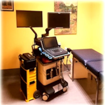
 Dr. Elena Venturini from San Raffaele Hospital in Milan discussed initial findings from the SOLUS project (Smart OpticaL and UltraSound diagnostics of breast cancer), showing that the system has promise for improved breast cancer diagnosis. The system incorporates optical methods, a smart optode performing diffuse optical tomography, and conventional ultrasound imaging, as well as advanced quantitative elastography. It uses the Aixplorer Mach 30 ultrasound system (Hologic) combined with a DOT system.
The European Institute for Biomedical Imaging Research (EIBIR) supports project management and leads dissemination activities. The project received funding from the EU's Horizon 2020 research and innovation program.
The prototype designed for the initial findings combines B-mode ultrasound, color Doppler, shear-wave elastography, and DOT through a probe that has a single ultrasound transducer in the center. The transducer is flanked by eight optodes that emit and receive light in the red and infrared spectrum. The probe is as big as a standard-sized 3D probe, and all of this is performed in one exam.
Eight wavelengths are used to collect data, which allows researchers to estimate tissue composition, as well as blood and scattering parameters.
For the pilot study, Venturini said the purpose was to test whether the method could characterize breast tissue, distinguish nodules from normal tissue, and determine whether lesions are malignant or benign. The SOLUS team also wanted to find out whether the method could be used while affecting clinical workflow as little as possible.
They tested the system on 24 women with breast nodules visible on standard ultrasound. Suspected lesions in the women were greater than or equal to 1 cm. A total of 12 optical acquisitions were taken from each patient.
The researchers found that the system has a sensitivity of 91% and specificity of 75%. Performance was assessed by machine learning algorithms based on lesion composition parameters gathered from optical data.
Venturini said the team's results need to be confirmed via a larger sample size. She added that one next step is to compare the method with other ultrasound methods, such as color Doppler and elastography, as well as further improve specificity.