Whole-lung radiomics features represent potential image biomarkers for improved fetal MRI-based prediction of abnormal lung development and neonatal respiratory function, researchers from the Medical University of Vienna have found in a new study.
Radiomics -- a quantitative image analysis method that is used to extract a large number of features from medical image data -- "is an exciting method that can help extract more information from images than is feasible by visual assessment alone," Dr. Florian Prayer, from the Department of Biomedical Imaging and Image-Guided Therapy, told AuntMinnieEurope.com.
"To take full advantage of radiomics, it is important to make sure that results are reproducible and the process of acquiring these results is transparent," he added.
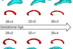
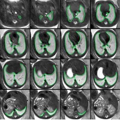
 Repeated acquisitions of T2-weighted axial MRI images of the lung in a fetus at gestational week 32. 3D regions of interest (ROIs) encompassing the whole lung were manually segmented in initial (green) and repeat (blue) acquisitions of 30 fetuses. 2D ROIs were defined as lung segmentations at the level of the carina in initial (red) and repeat (orange) acquisitions, respectively. Radiomics feature reproducibility was assessed between features extracted from 2 and 3D ROIs in the initial acquisition (red vs. green), 2D ROIs in repeated acquisitions (red vs. orange), and 3D ROIs in repeated acquisitions (green vs. blue). Figure courtesy of Dr. Martin L. Watzenboeck, Dr. Florian Prayer, et al, and Insights into Imaging.
Fetal MRI radiomics features extracted from 2D regions of interest (ROIs) do not adequately represent tissue characteristics of 3D ROIs encompassing the whole lung, the fetal imaging team in Vienna noted in an article posted on 8 February by Insights into Imaging. In addition, they exhibit insufficient reproducibility between repeated standardized fetal MRI acquisitions.
In contrast, a majority of fetal MRI radiomics features extracted from 3D whole lung segmentation masks are fully reproducible, the authors wrote.
Why the need for new data?
Currently, noninvasive in-vivo imaging assessment of fetal lung growth relies primarily on fetal ultrasound- or MRI-based lung volumetry, but this approach is limited by wide gestational age and body volume-adjusted normal lung volume ranges that may only inaccurately identify fetuses at risk for postnatal respiratory insufficiency, the researchers explained.
"Research on fetal MRI signal intensity-based analysis of lung maturity has produced variable results, thus far precluding its clinical translation," they noted.
To investigate the reproducibility of radiomics features extracted from 2-D ROIs versus whole-lung (3D) ROIs in repeated in-vivo fetal MRI acquisitions, the team carried out 30 fetal MRI scans, including two axial T2-weighted acquisitions of the lungs; 2D (lung at the level of the carina) and 3D (whole lung) ROIs were manually segmented.
The authors extracted a total of 95 radiomics features from 2 and 3D ROIs in initial and repeat acquisitions. Radiomics feature intra-class correlation coefficients (ICC) were calculated between 2 and 3D ROIs in the initial acquisition, and between 2 and 3D ROIs in repeated acquisitions.
They then assessed MRI data of 11 (36.7%) female and 19 (63.3%) male fetuses acquired at a median 25+0 gestational weeks plus days (interquartile range, 23+4 to 27+0 gestational weeks).
Median radiomics feature ICC between 2 and 3D ROIs in the initial MRI acquisition was 0.733 (interquartile range, 0.313-0.814; range, 0.018-0.970). ICCs between radiomics features extracted using 3D ROIs in initial and repeat acquisitions (median, 0.908 [interquartile range, 0.824-0.929; range, 0.335-0.996]) were significantly higher compared with 2D ROIs (0.771 [0.699-0.835, 0.048-0.965]) (p < 0.001).
Key findings
Overall, the authors are convinced the new study shows that radiomics can reliably maximize the diagnostic yield from fetal MRI studies. "Whole lung radiomics features are more sensitive to subtle differences between normal and pathological fetal lung development compared to 2D lung ROI features. Fetal MRI whole lung radiomics may improve non-invasive in-vivo lung development assessment," they stated.
Currently, the Vienna group is preparing a study that describes the radiomics signature of normal lung development in fetal MRI. "This will help to create a new image marker (observed-to-expected lung age) to identify fetuses at risk for neonatal respiratory insufficiency already during the second and third trimesters (weeks to months before birth)," Prayer explained.
The fetal imaging team will give several talks at ECR 2023, which begins on 1 March. Prayer's own presentation ("Foetal MRI radiomics predict gestational age based on foetal lung shape and texture") is scheduled for 4 March.