EuroMinnies' Finalists, Page 2
Most Significant News Event in European Radiology
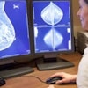
European Society of Breast Imaging recommends MRI for extremely dense breasts
After a long and protracted discussion among breast radiologists, the European Society of Breast Imaging (EUSOBI) finally recommended on 8 March 2022 that MRI be used for breast cancer screening in women with extremely dense breasts.


Published in European Radiology as an open-access article, the new recommendations marked a shift away from the one-size-fits-all approach of biennial mammography currently utilized in most European screening facilities in favor of personalized screening programs, according to the society.
EUSOBI said it arrived at its recommendations after carefully considering the evidence, particularly the Dutch Dense Tissue and Early Breast Neoplasm Screening (DENSE) trial.
"We have known for a long time that the performance of mammography screening in women with extremely dense breasts is not very good," said EUSOBI President Prof. Fiona Gilbert. "However, we had nothing better to offer. The recent MRI screening trials have largely changed this."
EUSOBI also called on all screening centers to provide women with their breast density information and for radiologists to inform women that there are also clear downsides of breast cancer screening. Women should have a say about how they want to be screened, according to Dr. Ritse Mann, PhD, chair of EUSOBI's scientific committee and first author of the European Radiology article.
"This implies on one hand that we must offer women all methods -- including breast MRI -- that are proven effective, while on the other hand, we should never hold it against them when they opt for less effective methods or even no screening at all," Mann pointed out.
Opposition to Ukraine war grows among Russian radiologists
 Street protests in Russia were relatively common after the invasion of Ukraine.
The Russian invasion of Ukraine on 24 February 2022 led to a wave of protests across Europe, even within Russia. By early April, nearly 18,000 people had signed the Russian physicians' petition, including many radiologists, according to Dr. Sergey Morozov, PhD.
Morozov, past president of the European Society of Medical Imaging Informatics, said that because speaking out against the war was a criminal offense in Russia, the signatories' names were obscured. He also noted that some medical doctors left Russia, but very few of them revealed the fact for fear of repercussions against themselves and their families.
On 30 March, Morozov resigned from his post as CEO of the Diagnostics and Telemedicine Center in Moscow and was then "removed from the office" of chief regional radiology officer of Moscow because of his antiwar posts on social media and his efforts to promote the physicians' petition. He left Russia and now works for the Belgian software developer Osimis as chief innovation officer.
A second respected radiologist who fled Russia earlier in March also spoke with AuntMinnieEurope.com. "Working in my hospital has been getting worse for several years, since the last election cycle. Everyone was pushed towards certain actions in favour of party power," he noted. "I had to resign."
The source continued: "I think the next few weeks could be devastating, to say the least. That horror has the potential to go on for an indefinite time. I am frightened, and I don't know how I am going to move to a safer place. The situation is so unnerving. I have to be really careful in my current situation, because it takes only one phone call and all could be over for me or my family."
Scientific Paper or Podcast of the Year
Can we increase efficiency of CT lung cancer screening by combining with CVD and COPD screening? Results of an early economic evaluation. Carina M. Behr et al, European Radiology, 1 January 2022
 Dr. Carina Behr.
This open-access article published online in European Radiology showed that combined low-dose CT (LDCT) screening for lung cancer, chronic obstructive pulmonary disease (COPD), and cardiovascular disease (CVD) can be cost-effective -- and it is likely to be more cost-effective than screening for lung cancer only.
Authors from the universities of Twente, Groningen, and Melbourne explained how chest LDCT, used in lung cancer screening, can detect early stages of COPD through emphysema or air trapping evaluation, and high CVD risk based on coronary calcium scoring.
They also revealed that to be cost-effective with the same screening population and willingness-to-pay (WTP) threshold, lung cancer-only screening should cost less than 113 euros per screened individual. However, this is only an early economic evaluation, and more knowledge into the co-occurrence of these diseases is required to identify the optimal target screening population, they emphasized.
The benefits of LDCT screening of COPD and CVD are not fully clear, but the authors believe the additional screening for these diseases within a lung cancer screening program could further improve outcomes at marginal additional costs as lung cancer screening patients often have high but unrecognized CVD risk.
"To our knowledge, this study is the first to apply a headroom analysis estimating the maximum acceptable cost for a screening program," noted first author Dr. Carina Behr, a doctoral candidate in health technology assessment at University of Twente, Enschede, the Netherlands, and colleagues.
The vanishing radiologist with Dr. Adrian Brady. Radcast episode by Drs. Uzoma Nnajiuba and Jamie Howie, 6 May 2022
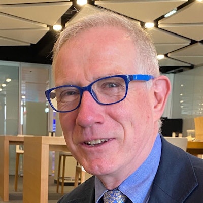
 Dr. Adrian Brady.
The commoditization of radiology endangers the capacity of the specialty to maintain its hard-won central role in patient care. This is the view of Dr. Adrian Brady, consultant radiologist at Mercy University Hospital in Cork, Ireland, and president of the European Society of Radiology and ECR 2023.
"To young radiologists I would say: make sure that you are part of the clinical team, that you are seen, and that you are a clinician first -- and a radiologist to support that clinical activity," he said in the Radcast episode.
Brady also elaborated on his previous comments about how technological improvements have led to the phenomenon of the "vanishing radiologist." Advances in CT and other modalities have increased reporting workload, and digitization of reporting has created the expectation of almost-instantaneous report availability, all of which reduces the time radiologists spend with referrers and patients, he noted.
Workload may in part be offset by AI tools taking on repetitive time-consuming tasks, such as looking for CT lung nodules, microcalcifications in breast imaging, or polyps in CT colonography. AI will probably also influence radiologists' work in other ways, but it will not make radiologists redundant, according to Brady.
"A practice that has taken 120 years to develop as a specialty won't be reduced to 1s and 0s or algorithms. A few years ago, medical students were asking if they should bother considering entering radiology as a specialty. Now they are no longer asking me that," he said.
Best New Radiology Device
Compact System 5000 Series ultrasound, Philips
Philips unveiled the Compact System 5000 Series at RSNA 2022. The portable ultrasound scanner is designed for shared service across specialties, including cardiovascular, obstetrics and gynecology, point-of-care, and general imaging.
 With the added battery, the Compact System 5000 Series gives users the flexibility to move between patients more quickly.
The device can be configured with an optional battery that allows for 2.5 hours of scanning time, and it features 33% fewer hard keys on the console and a fully sealed control panel and includes AI automation tools and support for its Collaboration Live real-time telemedicine software, according to the vendor.
Compatibility with Philips ultrasound systems Affiniti and EPIQ transducers, user interface, workflow, and automation tools allows an easy learning curve for advanced and novice users, the company added.
"We built the Compact System 5000 Series to perform as a highly versatile portable ultrasound system to be used in multiple care settings for many different types of exams without compromising image quality, diagnostic ability, or clinical workflow," said Jeff Cohen, general manager of Ultrasound at Philips.
"We find the system to be extremely user-friendly, particularly as it matches that of our EPIQ Elites we also have in our clinic. With the added battery, it also gives us the flexibility to move between patients more quickly," said Dr. Marcela Böhm-Vélez of Weinstein Imaging Associates, Pittsburgh, U.S.
Omni Legend PET/CT system, GE HealthCare
GE HealthCare launched the Omni PET/CT platform and Omni Legend system in October 2022 at the annual meeting of the European Association of Nuclear Medicine in Barcelona, Spain.
 At the Rambam Health Care Campus in Haifa, Israel, the Omni Legend has helped to increase throughput by over a third and cut dose by 40%.
The system uses an AI-based Auto Positioning Camera and Precision DL, a deep-learning image processing software, and it can scan beyond FDG with short-life tracers for cardiac and neuroimaging that can enable different procedures such as the diagnostics portion of theranostics imaging, the company added.
The device's Q.Clear PET image reconstruction software reportedly helps to ensure reliable quantification, while the MotionFree technology can correct respiratory motion artifacts for all patients, according to the vendor. Other features are a fast data quality assurance process for better calibration and simplified protocol selection on the gantry touchscreen and a new user interface.
GE HealthCare states that Omni Legend 32 cm increases small lesion detectability by 16% on average and up to 20%, as compared with the Discovery MI 25 cm with matched scan time/injected dose, as demonstrated in phantom testing using a model observer with 4 mm lesions.
At the Rambam Health Care Campus in Haifa, Israel, the product has helped to increase throughput by over a third due to shorter scan times -- achieving up to 35 patient scans during a 9.5-hour shift -- and reduce dose by 40% versus the old system, noted John Kennedy, PhD, chief physicist in the Nuclear Medicine Department.
Best New Radiology Software
Auto Scan Assist MRI software, Canon Medical Systems
Auto Scan Assist applies automation to the process of planning and performing scans of the brain, spine, cardiac, and knee, and uses deep learning and machine learning for liver, whole-spine, and prostate studies. It helps to standardize and simplify MR scan planning with automated slice alignment, and internal data show the burden on the operator can be reduced by up to 80% due to fewer operating steps and fast scan speeds, Canon stated.
 AI automatic positioning techniques allow staff to concentrate on other, more rewarding tasks, such as postprocessing.
Automatic slice orientation and positioning for sagittal, coronal, and axial planes in the cervical, thoracic, and lumbar regions is available for whole spine exams. Users can select a more detailed and exact slice orientation and positioning parallel and/or perpendicular to urethra for prostate examinations, according to the vendor.
In cardiac exams, the software can automatically detect 14 different standard views of the heart for quick workflow, while KneeLine+ improves reproducibility and image quality and supports the alignment of the knee to the iso-center, which reduces artifact-related rescans, it added.
"After 2 or 3 days of adaptation, the team has utmost confidence in the AI automatic positioning techniques, which allow them to concentrate on other, more rewarding tasks, such as postprocessing. Exam time is optimized and no time is wasted. Today, 85% of knee exams are performed using this technique," said Frédéric Martin, referring MRI technician at GIE Var Ouest IRM Scanner, Ollioules, France.
The software is offered on the Vantage Galan 3T, Vantage Orian, Vantage Fortian, and Vantage Elan, but not all features are available on the Vantage Elan, the company noted.
Deep Resolve Boost for MRI reconstructions, Siemens Healthineers
Deep Resolve Boost is a k-space-based reconstruction protocol that focuses on artifact reduction. It is a recent addition to the Deep Resolve AI-based reconstruction protocol for MRI, which was first introduced at RSNA 2020.
 The software is available on the Magnetom Vida MRI scanner.
Deep Resolve Boost enables users to acquire MRI exams with shorter time periods, such as a 15-second knee exam, with images then boosted to full image quality using the protocol, according to Siemens. Another protocol, Deep Resolve Swift Brain, enables a brain scan to be performed in less than two minutes.
Deep Resolve Boost is applicable from head to toe, and the "denoising strength" can be adapted by choosing from three levels. It can be combined with Deep Resolve Sharp and SMS TSE, rendering this the first k-space-to-image-space deep learning reconstruction that can be combined with simultaneous multi-slice turbo spin echo (TSE) imaging, the vendor claims.
"Image quality in standard 2D MRI sequences, accelerated in simulation beyond the threshold of standard acceptable noise levels, can be substantially improved by applying an iterative denoising algorithm using supplementary information about the image noise level," stated Dr. Johan Dehem, from Jan Yperman Ziekenhuis, Ypres, Belgium, in a Siemens-supported article about initial clinical experiences of the technology.
The software is available on the Magnetom Lumina, Magnetom Vida, Magnetom Vida Fit, Magnetom Altea, Magnetom Sola, and Magnetom Sola Fit. The minimum software version required is the Syngo MR XA50A and Syngo MR XA51A. Additional technical prerequisites may apply, stated the vendor.
Best New Radiology Vendor
CancerCenter.AI, Wrocław, Poland
CancerCenter.AI was co-founded in 2017 by Piotr Krajewski, who now works as the chief executive officer. He is an engineer and tech entrepreneur who also founded stermedia.ai, a provider of robotics, software, and design AI solutions to businesses.
 Piotr Krajewski (center) participated in a panel discussion at the "Made in Wrocław: Driven by Intelligence" conference in October 2021. The event is organized every five years by the City of Wrocław and Wrocław Agglomeration Development Agency.
"Our vision is to foster an ecosystem of apps and algorithms connected by APIs [application programming interfaces], of which we aim to be the gatekeepers," he said in an April 2022 interview with insightscare.com. "Data will be owned by the patient and available to doctors in accordance with GDPR and other medical Information Governance regulations. Our aim is to make high-quality cancer diagnosis available anywhere in the world, regardless of whether you live in a rich or poor country."
CancerCenter.AI supports oncology diagnosis using AI, deep learning, and machine learning. It aims to help physicians work together to detect cancer faster and more efficiently by using software platforms to diagnose patients without the necessity to deliver nondigital images.
Specialized algorithmic solutions analyze medical images for pathology and radiology. They are designed to offer rapid access for second and third diagnostic opinions by medical professionals, reducing anxiety caused by not knowing what they are dealing with and what is the exact nature of their disease, the company notes on its website.
"Our solutions do all the work -- segment images, find regions of interest, generate statistical descriptions of images, count cells, mitosis, and even recognize the type of cell. Your focus is saving lives, our focus is image processing and fast detection," it states.
Gleamer, Paris
Gleamer is a French medtech company developing AI for radiology. It was founded in 2017 by Christian Allouche, the chief executive officer; Alexis Ducarouge, the chief technical officer; and Dr. Nor-Edinne Regnard, the chief medical officer and a musculoskeletal radiologist. The firm now has over 50 staff and customers in 22 countries, mainly Europe, the U.S., and Australia.
 Gleamer's three co-founders (from left to right): Alexis Ducarouge, Nor-Edinne Regnard, and Christian Allouche. Photo courtesy of Jonathan Moyal.
Its main product, BoneView, enables radiologists, orthopedic surgeons, emergency physicians, rheumatologists, GPs, and physician assistants to diagnose fractures and traumatic injuries on x-rays. It received the CE mark class 2a certification in March 2020 and clearance from the U.S. Food and Drug Administration in March 2022.
At RSNA 2022 in Chicago, a BoneView clinical validation paper won the Alexander Margulis Prize for Scientific Excellence. Published in Radiology on 21 December 2021, this study used multireader, multicase methodology based on an external multicenter data set of 480 exams with at least 60 examinations per body region from July 2020 to January 2021.
Lead author Dr. Ali Guermazi from Boston University School of Medicine concluded that AI assistance improved the sensitivity and may even improve the specificity of fracture detection by radiologists and nonradiologists, without lengthening reading time. Regnard was a co-author.
In the first quarter of 2023, Gleamer plans to launch two new solutions for the automation of the measurements and the automation of the bone age assessment. It also hopes to publish more clinical studies in major journals.