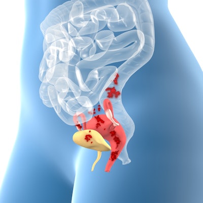
Ultrasound detected signs of endometriosis in approximately one-third of young women presenting with severe dysmenorrhea, according to Italian research published online on 5 December in Fertility and Sterility.
A team led by Dr. Francesco Martire from the University of Rome found that accurate pelvic ultrasound scans help provide early diagnosis in these women by identifying small endometriotic lesions.
"Young patients with dysmenorrhea should be referred to an expert sonographer to minimize the delay between the onset of symptoms and diagnosis," Martire and colleagues wrote.
Endometriosis is typically diagnosed later in life and affects only about 10% of premenopausal women worldwide, reports say. Most patients are referred with dysmenorrhea in adolescence or at a young age at or below 25 years.
However, the researchers noted that this symptom is underestimated and considered a normal symptom in young women. Along with that, clinical and pelvic exams typically yield negative results, leading to potential missed early diagnosis. With this in mind, the researchers suggested that the true prevalence of endometriosis in young women remains unknown.
Laparoscopy is the gold standard for diagnosing endometriosis, but some clinicians may be hesitant about using this invasive technique. Noninvasive ultrasonic imaging has been explored in this area, with the European Society of Human Reproduction and Embryology recently recognizing imaging as the standard for endometriosis diagnosis.
Martire and his colleagues aimed to explore the presence of different endometriosis forms, either isolated or combined, using ultrasound images in women aged 25 years or younger. These women presented with dysmenorrhea. The group also wanted to assess other painful symptoms linked to endometriosis or adenomyosis and how they appear on ultrasound findings.
The study included 371 women aged 12 to 25 years who were referred between 2016 and 2021 with severe dysmenorrhea and a visual analog pain scale of 7 or above. The researchers used 2D, 3D, and power Doppler pelvic ultrasound on all patients.
They found that at least one ultrasound endometriosis feature was identified in 131 (35.3%) patients. Ultrasound, however, was normal in 170 (45.8%) women despite the referred dysmenorrhea, with the normalcy rate being 69.2% in the age 12 to 15 subgroup. This was statistically significantly different from the other age subgroups.
With respect to ultrasound findings, endometriosis presented in just 12.8% of women between the ages of 12 to 16, compared with 42.6% seen in women ages 21 to 25.
Of the 131 patients with endometriosis, ovarian endometrioma was found in 54 (41.2%), with 22 (16.8%) having an isolated endometrioma. Additionally, adenomyosis was detected in 67 (51.1%) patients, and 28 (21.4%) showed its isolated indications. Posterior deep-infiltrating endometriosis was also found in 70 (53.4%) patients, and uterosacral ligament fibrotic thickening was found in 63 (48.1%). The uterosacral ligament was completely isolated in 23 women.
Finally, the researchers also found that the combined occurrence of dysmenorrhea with dyspareunia, bowel symptoms, and heavy menstrual bleeding increases the presence of endometriosis up to 59%, 63%, and 45%, respectively.
The study authors wrote that these results highlight the importance of early noninvasive diagnosis of endometriosis in these women by combining symptoms with pelvic ultrasound exams.
"Isolated disease findings, mostly small uterosacral ligament thickening, mild adenomyosis, and small endometriomas, are common features of endometriosis associated with severe dysmenorrhea in young patients," they added. "These results are consistent with endometriosis probably starting early in life."