Over the last year, I have helped to install several low-field MRI systems and seen many more being installed at other sites. It has been remarkable to watch an MRI system simply wheeled out of its crate and into the room where it is going to be used.
While the physical installation process is quite different from that of a high-field system, many of the requirements still exist, such as IT infrastructure and trained staff to operate the scanner. Local rules within the facility might also have to be adapted to accommodate a portable MRI system, where the safety zones are no longer static but move with the scanner.
Another aspect of safety is scanning patients with implants or active devices. The lower field strength will reduce specific absorption rate (SAR) down to minimal levels, and reduce attractive forces, but there currently are limited guidelines for imaging implants in vivo at low field.
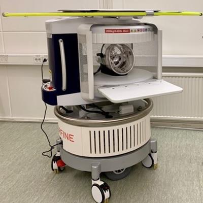
 Emil Ljungberg holds the tablet that controls the 64 mT Hyperfine scanner.
An exciting opportunity offered by low-field MRI is the greatly reduced cost by using a permanent instead of superconducting magnet. The reduced field strength makes it possible to use the scanner in a non-shielded room, by instead using active noise-cancellation and a radiofrequency (RF) shield.
Reducing the system and siting cost directly lowers the threshold for introducing MRI in new settings, such as smaller hospitals and healthcare clinics with limited budgets, and crucially opens the door to widening global access to brain imaging.
Historic drive to high-field
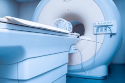
Since the discovery of in vivo MRI, there has been a race towards higher field strengths in search of better images. At Lund University, we have experienced this development firsthand, from the installation of a 0.07-tesla scanner built in the early 1980s to a 7T system installed in 2015. But the push for a higher field strength comes at a cost, both in monetary and logistical terms. High field MRI scanners are expensive to manufacture and maintain and require dedicated hospital infrastructure, which can contribute to reduced access to MRI.
 The first MRI system built in Lund, Sweden, in the early 1980s, along with its inventor Prof. Emeritus Bertil Persson. (Courtesy of Persson, BRR. and Ståhlberg, F. 2017. Så började klinisk NMR i Lund. Acta Scientiarum Lundensia, 2017-001, pp.1-15. ISSN 1651-5013).
In parallel with the field-strength race, great advancements have been made in system design and image reconstruction algorithms, both of which have contributed to improved image quality. These technologies have now made it possible to look in the rear-view mirror and revitalize low-field MRI as a cost-effective and portable supplement to high-field scanners. Many research institutions have demonstrated various designs for low-field MRI systems (~50 mT), and in 2020, Hyperfine, a U.S. MRI manufacturer, received Food and Drug Administration clearance for its low-field, portable MRI system for clinical use.
The renewed interest in low-field MRI is driven by unique aspects of low-field systems which can improve the value of MRI in many ways. Earlier this year, we installed the 64mT Swoop system, thus taking us full circle, back to where MR research started here in Lund.
Added value of portable low-field MRI
The return to low-field MRI is not a return to the image quality of the first MRI scanners from the 1980s. We tend to associate the field strength with scanner performance, but the magnet is but one component.
Thanks to the developments of high-field system technology over the last decades, it is now possible to produce clinically meaningful images at low field (<0.1T).1 However compared to modern high-field scanners, low-field MRI will always pay a penalty of lower signal to noise ratio. To fully appreciate the opportunities low-field MRI offers, we need to expand our view of the value added by MRI.
In a standard radiological workflow, patients are transported from the ward to radiology for imaging, which can introduce considerable risks. Low-field MRI systems can be made portable and thus reverse the workflow by bringing the scanner to the patient, a concept realised with portable ultrasound, x-ray, and CT scanners. One direct application of low-field MRI is stroke imaging where a portable system taken to the patient can reduce the time to imaging, which is crucial for deciding on appropriate treatment.1
A common cause of reduced access to MRI is contraindications such as implants or other non-MR conditional devices. Reducing the field strength will reduce these risks and enable imaging of a wider patient cohort. However, as low-field MRI is re-emerging as a new category of scanners, evidence and guidelines for safety limits are still in development, and MRI safety will always be the primary focus.
Worldwide access
The IMAGINE project of the International Atomic Energy Agency (IAEA) has gathered data on the availability of medical imaging technology around the world, and its results paint a sobering picture. There are vast disparities in the number of MRI scanners globally, with over 100 times more MRI scanners in high-income nations than low-income countries. More so, a single number does not convey how well these systems can be accessed by the general population. It is expected that low-field MRI systems can help close this resource gap and deliver brain imaging to the global population.
In 2021, we embarked on a research project funded by the Bill and Melinda Gates Foundation, led by Prof. Steven Williams at King's College London in collaboration with Hyperfine, to investigate the utility of low-field MRI in regions that previously have had poor access to MRI. Some 20 sites are involved in the project, a combination of physics and engineering groups focusing on pulse sequence development, together with clinical partners to integrate low-field MRI in clinical studies. It is a world-wide project spanning from Canada to Pakistan, from Sweden to South Africa.
This is the first study to investigate the utility of low-field MRI on a global scale, so we are all excited to see the potential of low-field MRI being realized.
Will low-field replace high-field?
It is probably evident at this point that I am a strong proponent of the development and introduction of low-field MRI in research and clinical practice, but I do not see low-field MRI replacing our high-field systems.
With portable low-field systems, we are no longer tied to the radiology suite, and we should consider how low and high-field systems are best used together. With more options come increased possibilities of combining high and low-field imaging for improved outcomes and added value to the patient. This initiative will only succeed through collaboration between clinicians who know the clinical need and imaging scientists with the skills to further develop the technology.
Increased access to MRI examinations does not, by itself, guarantee increased access to diagnostic results. If the number of exams increases without commensurate growth of the radiology staff, we only move the bottleneck in the system. We need to consider feasible methods for reading an increasing number of scans produced by low-field systems, such as artificial intelligence (AI)-assisted techniques.
I am eager to see the scope of MRI research expanding across field strengths. Perhaps we will break free from the field strength race and consider the optimal field strength for a given application. Advancements made at high field can be utilized at low field and vice versa, thus driving the field forward together.
The MRI research field has always maintained a high pace, and now we seem to finally have reached the speed needed to take us back to the future and reinvent low-field MRI.
Emil Ljungberg, PhD, is an MRI physicist in the MR Physics Group at Lund University, Sweden, and a visiting research fellow at the Department of Neuroimaging at King's College London. His research focuses on development of novel imaging methods at low field, as well as acoustically silent neuroimaging using zero echo time (ZTE) MRI.
The comments and observations expressed herein do not necessarily reflect the opinions of AuntMinnieEurope.com, nor should they be construed as an endorsement or admonishment of any particular vendor, analyst, industry consultant, or consulting group.
Reference
- Mazurek, M. H. et al. Portable, bedside, low-field magnetic resonance imaging for evaluation of intracerebral hemorrhage. Nat. Commun. 12, 1–11 (2021).