The clinical benefits of photon-counting CT are becoming clear and obvious in both cardiovascular and thoracic imaging, according to a leading developer of this emerging technology.
 Thomas Flohr, PhD. Courtesy of Deutscher Zukunftspreis/Ansgar Pudenz.
Applications in these two areas are mostly likely to drive its wider introduction in hospitals because of its many technical advantages over conventional CT, Thomas Flohr, PhD, said in a keynote lecture at the British Institute of Radiology (BIR) annual congress in London. He noted that surgeons particularly like the images because they show the vessels in much more detail than they are used to seeing.
"Photon-counting CT can also open up the potential to use coronary CT angiography (CTA) as a first-line test for patients with recurrent chest pain after stenting. This is a large patient group that today mostly has to be excluded," said Flohr, who has worked as head of CT physics and application predevelopment at Siemens Healthineers since 2004.
In the Sir Godfrey Hounsfield memorial lecture at the BIR congress on 22 September, Flohr predicted that in time, system costs of photon-counting CT will fall, saying there was "no fundamental limit which will prevent the cost of these systems from coming down."
Why the need for photon-counting CT?
Flohr, who is an adjunct professor for medical physics at Eberhard Karls University of Tübingen, Germany, described how CT had progressed over time since Hounsfield's brain scanner but had hit limits to further expansion. For example, spatial resolution was insufficient for demanding tasks, such as coronary CTA, and it was not recommended for patients with stents and recurrent chest pain.
Moreover, CT radiation dose remained a concern and findings "frequently lacked clinical significance," so follow-up examinations were needed for clear diagnosis and decisions on clinical management, he told BIR delegates.
By comparison, photon-counting CT offered clear advantages by using a new kind of detector that differs from a standard energy integrating detector (scintillator). Photon-counting detectors directly transform x‐ray photons into electrical signals (without the two-step conversion process in a scintillator), to produce a digital signal that is not affected by electronics noise, explained Flohr, whose team was nominated for the 2021 German Future Award.
He said photon-counting CT could be "twice as sharp" as a standard scintillator CT because the detector pixels can be made smaller, while offering improved contrast to noise ratio, at lower radiation dose and with intrinsic spectral information.
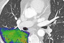
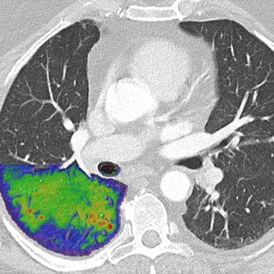
 Lung images of a post-COVID patient with photon-counting CT. Photon-counting allows for simultaneous acquisition and visualization of detailed structures (middle image) combined with functional information (image on the right). For comparison, an image acquired with conventional CT technology (on the left). Courtesy of Prof. Dr. Jiří Ferda, PhD, University Hospital Plzen, Czech Republic.
Research had shown that in thoracic imaging, it not only provides sharper images but also could "increase reader confidence" for a diagnosis of interstitial lung disease, he added.
"Of course, further clinical studies will be needed to demonstrate the clinical potential photon-counting has, in differential diagnosis and follow up of lung diseases," Flohr said.
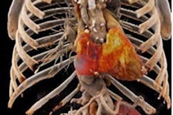
Clinical promise in cardiac imaging
Good spatial resolution was also "of the essence" in cardiovascular imaging.
"The biggest risk factor to obtain a non-diagnostic coronary CTA is still the presence of severe calcifications," he continued. "With its high spatial resolution, photon-counting CT can address this challenge. The overestimation of stenosis because of calcifications can be significantly reduced with photon-counting CT."
Research had shown that the diagnostic quality of images obtained with the technology "was much better with regard to calcium blooming" compared with energy-integrating CT.
The quantification and characterization of coronary plaques is another hot topic in cardiology today because both the total plaque burden and the composition of the plaques are an indicator of the likelihood of future cardiac events. And plaques with a high lipid content are particularly suspicious," he noted.
Photon-counting CT can help with improved plaque characterisation and quantification. He said spectral data, in conjunction with very high spatial and temporal resolution, could also enable new applications to make CT more reliable and more accessible to a wider patient range.
"I think we are likely to see that the great wealth of information in the high resolution spectral photon-counting data will be exploited even better with learning-based algorithms," he concluded.