Researchers at Aarhus University Hospital Skejby's radiology department have used CT and MRI to shine a light on one of the world's rarest fishes, the coelacanth. They say they've gained new insight into how it functions, and this knowledge may contribute to saving the critically endangered deep-sea dweller.
"This fish is iconic, extremely rare, and remains shrouded in mystery. It is difficult to observe alive because it lives in underwater caves at depths of 150-200 meters. And, since a large portion of the few specimens collected over the years have been cut to pieces, new methods were required to find out more about how it lives. We now know a bit more," said Peter Rask Møller, an associate professor and curator at the University of Copenhagen's Natural History Museum of Denmark.
There are two living species of coelacanth: Latimeria chalumnae, which lives off the coast of East Africa, and Latimeria menadoensis, which lives off Indonesia. The oldest known coelacanth fossils are more than 410 million years old. Just over 300 live specimens of coelacanth are believed to have been caught worldwide. Coelacanth are pregnant for five years and give birth to live young. Coelacanth grow up to two meters long and weigh up to 100 kg.
The Danish coelacanth specimen was caught in the Indian Ocean's Comoros archipelago in 1960. At the time, the Comoros was a French colony. Danish oceanographer Anton Bruun persuaded France to donate the specimen to the Zoological Museum in 1962.
With the donation came strict requirements that the specimen was only to be used as a display item and could under no circumstances be subjected to dissection. This requirement was unofficially lifted in 1975, when the Comoros gained independence from France, Copenhagen chose to honor the original agreement, so the specimen was kept intact. Over the years, the specimen has been exhibited at the Zoological Museum and the former Denmark's Aquarium.
Since it was first discovered in South Africa in 1938, many other coelacanth specimens have been dissected, so its anatomy is no secret, but very little is known about the fish's physiology.
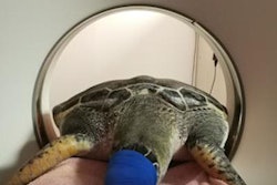
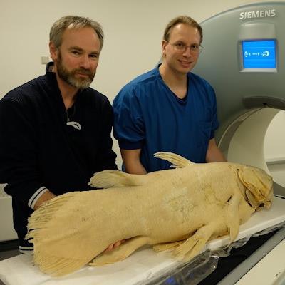
 Peter Rask Møller and Henrik Lauridsen with the Copenhagen coelacanth at the CT scanner, preparing the specimen for high-resolution imaging spanning an entire night. Image courtesy of Henrik Lauridsen.
By putting the fish in CT and MRI scanners at Aarhus University Hospital in Skejby, the research scientists could model the species with more precision than ever, while not destroying the fish. The models show the exact distribution of bone mineral and fat in its body. Among other things, the models help explain the unique "headstand drift hunting" technique of the coelacanth, whereby it slowly drifts along a seabed vertically, head and snout downwards, as it uses an electrosensitive organ to scan the bottom for cephalopods and fish to eat.
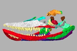
"We discovered that the coelacanth has a special skeleton with a lot of bone mass in the head and tail, while there are almost no vertebrae. It's quite unique. The heaviest parts are at either end of the fish, which makes it easy for the fish to stand itself on its head. The balance point is an advantageous mechanism for its way of life," explains Associate Professor Henrik Lauridsen from Aarhus University's Department of Clinical Medicine.
The researchers also discovered the precise distribution of fatty tissue in the body of the fish, including the amount in its fatty bladder, as coelacanth don't have a regular gas swim bladder like modern fish. The numbers show that the fat content correlates with the depths at which the fish live, where fat allows the fish to be neutrally buoyant and spend hardly any energy to remain hundreds of meters deep.
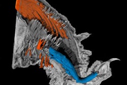
 Prey items in the gastrointestinal tract and prey with a swim bladder. (a-d) Mineralized remains of prey items in the distal portion of the digestive tract of the coelacanth imaged by CT and MRI. Dashed gray square in (a) is magnified in (b) and shown in a similar view plane using T2-weighted MRI (c). A 3D surface rendering of the fecal matter is shown in (d). Sagittal T2-weighted MRI slice (e) and three components (skin, bones, swim bladder) model (f) made from CT and MRI of Beryx decadactylus, a known prey item of the coelacanth. This species contains an air-filled swim bladder (light blue segment in f) that under the assumption of neutral buoyancy displaces a volume of seawater with the same mass as the net weight of the fish (excluding the swim bladder) in seawater. Image courtesy of Dr. Henrik Lauridsen and BMC Biology.
One peculiar feature about the coelacanth is that females gestate for a full five years before birthing live young. One of the great coelacanth mysteries among researchers is: Where do coelacanth give birth? The Danish researchers hope to shed light on this question soon.
"We still have no idea where their fry are born. By analyzing the distribution of bone and fat in a fetus, we can probably find out at what depth fry are adapted to live. This knowledge is also important in terms of preserving this critically endangered species -- because when we don't know where they are, we can't know where to protect them. And, there is cause for concern. Coelacanth have an incredibly slow reproduction rate, which makes them extra vulnerable," said Lauridsen.
The researchers point out that their models can be applied to all other aquatic organisms and used to determine, among other things, whether organisms are adapted to the ocean depths at which they live. This is relevant knowledge at a time when climate change could cause ocean currents to change and thereby impact marine life.
They now plan to use imaging on preserved specimens of juvenile coelacanths.
"They are born live after five years of gestation (the longest in the animal kingdom), but nobody knows at what depth," Lauridsen told AuntMinnieEurope.com. "By having bone mineral (CT) and fat tissue (MRI) content and distribution, we may be able to model what depth they are adapted to be neutrally buoyant, at which would be the expected depth of birth because a small fish cannot afford to fight gravity all the time."
Full details about the imaging technique used by the authors are available in the open-access paper, published on 19 August in BMC Biology.