Setting radiation doses on mammography systems manually can potentially reduce exposure when imaging thicker breast tissue. However, auto filtering is still the way to go in standard mammography practice, a study published on 19 August in Radiography found.
Researchers led by Devanshi Patidar from the Singapore Institute of Technology found reduced radiation dose for manual mode techniques used for 6 cm and 6.5 cm breast thickness in their phantom model. However, no significant differences were found in thickness measurement between 4.5 cm and 5.5 cm, and manual settings resulted in reduced image quality for some mammograms.
"For 4.5, 5.0 and 5.5 cm phantoms, as average glandular dosage was not significantly different among the different modes, the autofilter may remain the most practical option," Patidar and colleagues wrote. "However, significant reductions ... were obtained for the 6.0 and 6.5 cm phantoms when manual mode techniques were used."
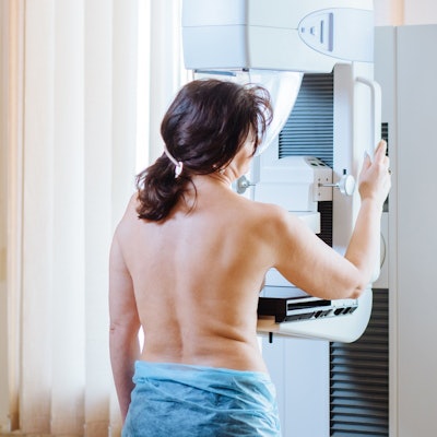
While digital mammography remains the gold standard for breast cancer screening and detection, some have expressed concern over the use of ionizing radiation in breast tissue.
Mammography units nowadays come with manual and automatic exposure modes. For manual modes, the operator can select and customize imaging parameters such as target and filter material, kilovoltage peak (kVp), and milliampere seconds (mAs). Mammography units can also automatically select exposure factors based on breast thickness and composition.
One such automatic mode, autotime, allows automatic mAs selection with manual target and filter material and kVp selection settings. Another, auto-filter, automatically selects all exposure parameters.
While autofilter has been the go-to option for screening mammography, recent research on phantom models suggests that there are tradeoffs in dosage and image quality when using either auto or manual controls for varying levels of breast thickness.
Patidar et al wanted to explore this idea themselves, looking at the effectiveness of autofilter, autotime, and manual exposure modes of a digital mammography system for radiation dose and image quality.
They used breast phantoms with levels of thickness ranging from 4.5-6.5 cm and had three mammographers independently analyze a total of 125 images. Polymethyl methacrylate blocks were used on the phantoms to reflect varying thicknesses. The researchers analyzed images with acceptable quality by comparing their average glandular doses.
| Comparison of average glandular doses (in mGy) for auto and manual exposure modes | |||
| Phantom thickness | Autofilter | Autotime | Manual |
| 4.5 cm | 1.2 | 1.2 | 0.92 |
| 5 cm | 1.38 | 1.38 | 1.17 |
| 5.5 cm | 1.68 | 1.71 | 1.27 |
| 6 cm | 2.03 | 1.98 | 1.52 |
| 6.5 cm | 2.33 | 2.38 | 1.86 |
The study authors wrote that statistical significance was achieved for dosage between the autofilter and manual modes for a thickness of 6.0 cm (p = 0.008), as well as between the autotime and manual modes for a thickness of 6.5 cm (p = 0.047).
The gaps in reported mAs used between auto and manual modes grew wider as thickness increased. For a phantom thickness of 4.5 cm, the autofilter and autotime modes used 80 mAs each, compared with 32 mAs used for manual mode. For a thickness of 6.5 cm, the autofilter mode used 137 mAs and the autotime mode used 191 mAs. Manual mode however used just 45 mAs.
Patidar and colleagues also noted that there was an "excellent" level of agreement among the mammographers. All images taken for the 4.5 cm, 5 cm, and 5.5 cm phantoms met the minimum criteria set by the American College of Radiology (ACR). However, 12% and 28% of the images taken for the 6 cm and 6.5 cm phantom respectively, failed to meet such criteria.
Using autofilter mode as a reference, just 50% and 61.1% of manual mode images met the image quality criteria for 4.5-cm and 5-cm phantoms, respectively. Even less manual mode images for thicker levels met the criteria.
The team concluded that autofilter mode speeds up the imaging process by reducing the time needed for manual selection of parameters and reduces the risk of operator error but that manual settings could help with imaging thicker breast tissue. The group called for further work to evaluate the appropriate use of these modes and added that their findings will support related future human breast studies.