The question today isn't why PET/MRI hybrid scanners should be used over PET/CT but how they can be best put to use. This is the view of a top German specialist in hybrid imaging, who points to four major applications in which PET/MRI is proving to be a game changer.
Several cornerstones have been embedded in the literature regarding the advantages of PET/MRI over PET/CT, which notwithstanding is a more established hybrid approach, Prof. Dr. Lale Umutlu, vice chair of radiology and head of clinical PET/MRI at University Hospital Essen, told ECR 2022 attendees. Those advantages include increased soft-tissue contrast, the value of integrated multiparametric MRI (mpMRI) and metabolic data, and a reduction in radiation exposure to patients.
"As far as clinical application fields of PET/MRI, they can be subdivided into 'The Big Four,' as I like to call them, and these are inflammatory diseases, cardiovascular diseases, neuroradiology, and oncological imaging," Umutlu proposed.
 Prof. Dr. Lale Umutlu presented images of the "Big Four" clinical applications for hybrid PET/MRI. All images courtesy of Prof. Dr. Lale Umutlu and ECR 2022.
1. Inflammatory diseases
At University Hospital Essen, PET/MR enterography is used in many cases because the clinical team has learned it improves specificity for detecting active inflammatory disease, not just in Crohn's disease but also in ulcerative colitis, she said.
"What you can do is perform a complete protocol of MR enterography, and simply add the PET component to better differentiate between scarring and fibrosis and active inflammation," Umutlu noted.
For the same reasons, PET/MRI is also proving to make a difference when it comes to imaging spondylodiscitis and large-vessel giant cell arteritis, she added.
2. Cardiovascular diseases
Cardiovascular imaging with PET/MRI is challenging because it is susceptible to artifacts and patient preparation can be difficult, as dietary restrictions are stricter than conventional FDG imaging, Umutlu said.
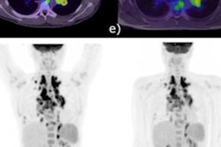
However, in compliant patients, PET/MRI can be used to diagnose acute myocardial infarction, where MRI provides late enhancement images suggesting myocardial hypometabolism and PET FDG radiotracer uptake further allows the differentiation between healthy tissue and scarring.
 Hybrid late-enhancement MRI and PET imaging can help diagnose acute myocardial infarction.
PET/MRI can also be used to diagnose acute myocarditis, and in fact, in some cases has improved diagnostic confidence when infections aren't clear on MRI, she continued. This again is due to the addition of PET radiotracer uptake in the images showing active inflammation.
3. Neuroradiology
Clinical use of PET/MRI is most firmly established in neuroradiology at University Hospital Essen, partly because it's easier and faster than cardiovascular imaging due to less movement and a smaller field of view.
"Our neurosurgeons like the PET/MRI scan for assessing where to perform the biopsy in terms of highest [radiotracer] uptake," she said. In other words, areas of brain tumors with the highest tracer uptake indicate the most aggressive part of the tumor and therefore can improve biopsy results.
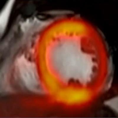
Also, neuro-oncologists use PET/MRI to monitor patients undergoing treatment, primarily to identify glioblastomas that may have returned after surgical removal. Umutlu showed an example of such a case in which MRI alone would probably have missed a tumor, but that was clearly shown with the addition of PET.
 The PET component of PET/MRI can reveal brain tumors that may go undetected by MRI alone.
"This was one of the cases where the PET component really helped us make a clear diagnosis in terms of a second manifestation of a brain tumor," she said.
4. Oncologic imaging
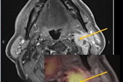
In Essen, PET/MRI is used most robustly in oncologic imaging, specifically prostate imaging. PET/CT can and is used effectively for detecting biochemical recurrence after prostatectomy, but prior to surgery, PET/MRI is superior for detecting primary tumors as well as cancer that has spread to surrounding tissue.
"This is one of the clearest application fields actually for PET/MR," Umutlu pointed out.
 PET/MRI is significantly more effective than PET/CT for imaging prostate cancer prior to surgery, Umutlu said.
In addition, PET/MRI is being applied successfully in soft-tissue sarcoma imaging, neuroendocrine tumor imaging (gallium-68 DOTATOC-PET/MRI), and in whole-body staging, where PET/MRI is equal to PET/CT in terms of tumor detection and acquisition times, yet holds an edge in classifying additional findings that may be indeterminate of PET/CT.
Ultimately, while clinicians continue to use PET/MRI more frequently and successfully in these "big four" areas, researchers are moving forward to integrate hybrid imaging data in radiomics and artificial intelligence applications, according to Umutlu.
In one study, researchers tested multiparametric MRI/PET imaging data (a virtual biopsy) compared with invasive biopsy and histochemistry for diagnosing brain tumors, and they found the virtual biopsy to be just as effective.
"I believe this will probably change our future," she concluded.
While the modality's share of the medical imaging market remains small, the global PET/MRI market is estimated to approximately double by 2029, according to market researchers. Since PET/MRI's introduction 10 years ago, more than 110 scanners have been installed worldwide, with 23% in North America and 54% in Europe.