Artificial intelligence (AI) trained with ultrasound images can help differentiate between malignant lymph nodes from benign nodes that react as a side effect of COVID-19 vaccination, found researchers in a study published on 7 July in the European Journal of Radiology.
Researchers led by Dr. David Coronado-Gutiérrez from the Hospital Clínic de Barcelona in Spain found that their method, which retrained an old machine-learning algorithm with ultrasound images, outperformed radiologists in determining which nodes were malignant or benign.
"These techniques could be useful to non-invasively diagnose lymph node status in patients with and without diagnosed breast cancer with suspicious reactive nodes," Coronado-Gutiérrez and colleagues wrote.
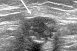
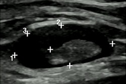
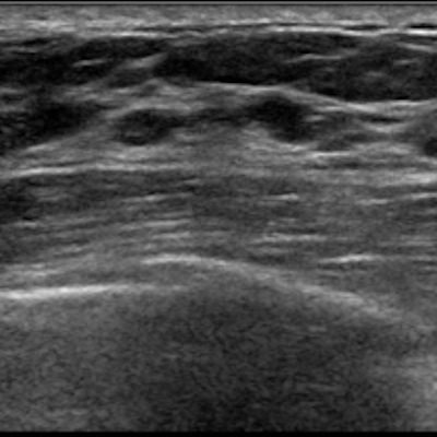
Swollen lymph nodes -- otherwise known as lymphadenopathy -- are a known, yet less common side effect of the COVID-19 vaccines. Swollen nodes can appear on imaging, causing concern and anxiety for patients who may think they are a sign of breast cancer. Radiologists and oncologists have been advised to be aware of the vaccination status of patients so that they aren't recommended for unnecessary follow-up or biopsy.
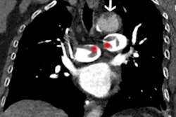
Ultrasound is used to assess lymph nodes before breast cancer surgery. Invasive techniques such as core-needle biopsy and ultrasound-guided fine-needle aspiration are used to confirm malignancies for suspicious nodes.
The study authors have explored the potential of AI to noninvasively predict axillary lymph node involvement in breast cancer from medical images, and the Coronado-Gutiérrez team previously studied AI methods in diagnosing metastatic involvement in axillary lymph nodes from ultrasound images.
In the group's most recent study, the researchers wanted to find out the potential of similar AI techniques and image analysis in differentiating between malignant lymph nodes affected by breast cancer from benign nodes caused by COVID-19 vaccination.
The team recruited a total of 180 new images from 154 different patients. Out of these, 71 images (10 cases and 61 controls) helped retrain the team's old model. Another 109 images (36 cases and 73 controls) were used to evaluate the retooled model's performance.
| AI model vs. radiologists in differentiating between malignant and benign swollen lymph nodes after COVID vaccination | ||
| Radiologists | AI model | |
| Accuracy | 69.7% | 92.7% |
| Sensitivity | 41.7% | 77.8% |
| Specificity | 83.6% | 100% |
The authors wrote that this technique could be used in clinical practice as an online tool or integrated into an ultrasound scanner. They added that radiologists can use the algorithm right after exploring an axillary lymph node, with results being obtained in less than 30 seconds.
"This software could be used to support decision making in both cancer and non-cancer patients with suspicious lymph nodes during ultrasound examination," they wrote. "In this way, unnecessary biopsies would be avoided, thus improving patient management and treatment."
The researchers also touted that this method could also be helpful after neoadjuvant chemotherapy by noninvasively evaluating axillary lymph node status. This in turn can help avoid unnecessary excisions.
The team also called for larger, multicenter studies to confirm and validate their results.
"In the future, if larger databases were collected, an automatic node's delineation could probably be implemented using deep learning algorithms given their recent success stories in similar applications," Coronado-Gutiérrez and colleagues wrote.