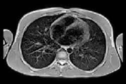
A novel MRI technique using hyperpolarized xenon-129 gas was able to illuminate lung abnormalities in patients with long COVID-19, even those who were not admitted to the hospital, according to a U.K. study published on 24 May in Radiology.
Called Hp-XeMRI, researchers using the technique found persistent lung abnormalities not seen on CT or chest x-ray, said a group led by James Grist, PhD, of the University of Oxford.
"Using Hp-XeMRI may enable us to further understand the cause of breathlessness in long COVID patients, and ultimately lead to better treatments to improve this often-debilitating symptom," Grist said in a statement released by the RSNA.
The Oxford team's latest publication follows the initial report on the technique covered by AuntMinnieEurope.com in article posted on 1 February 2022.
People who have contracted COVID-19 may experience symptoms and effects long after the acute phase of the illness, including breathlessness, fatigue, and brain fog, the investigators noted. But Hp-XeMRI can identify abnormalities of alveolar gas exchange that may not appear on CT scans or chest x-rays.
"These abnormalities are not apparent on conventional imaging, and in some individuals were detected up to a year after their initial COVID-19 infection," senior author Prof. Fergus Gleeson, head of academic radiology, director of the Oxford Imaging Trials Unit, and divisional director for the Oxford University Hospitals NHS Trust, said in the RSNA statement.
The investigators conducted a study that included 11 individuals with long COVID who had not been hospitalized during the acute stage of the illness and 12 people who had been admitted to the hospital for the disease; all had symptoms of breathlessness. The nonhospitalized group was 240 to 334 days past initial infection, while the posthospitalized group was 105 to 190 days past initial infection. The study also included a cohort of 13 healthy individuals who had no evidence of prior COVID-19 infection.
Study participants who had had COVID-19 underwent chest CT, Hp-XeMRI, pulmonary function tests, and one-minute sit-to-stand tests, while the healthy controls underwent only Hp-XeMRI.
The study found "significant differences in the mean red blood cell to tissue plasma ratio between healthy controls and posthospitalized COVID-19/nonhospitalized long COVID participants, indicating potential differences in lung function," and that "nonhospitalized long-COVID participants had near-normal CT scores, and [gas transfer, or DLco (%)] was significantly lower between nonhospitalized long-COVID and posthospitalized COVID-19 participants, potentially indicating a decrease in lung function but not structure," the team reported.
The findings may help clinicians and their patients get a better handle on a complicated disease, wrote Grace Parraga, PhD, and doctoral candidate Alexander Matheson, both of Western University in London, Canada, in an accompanying editorial.
"The COVID-19 pandemic has provided unprecedented challenges and important opportunities to better understand the natural history of viral lung infection in millions of patients," they wrote. "These hyperpolarized 129Xe MRI findings reveal new valuable clues about lung abnormalities that endure, months post infection, perhaps putting post-COVID patients back in the driver's seat of their recovery."