Researchers from University Hospital Cologne have reported in a proof-of-concept study that two PET/CT radiotracers -- F-18 FDG and gallium-68 FAPI-46 -- work better together than when used alone for cancer imaging.
The group injected both tracers prior to PET/CT imaging in a small group of patients diagnosed with head and neck cancer. Clinical experts visually detected more suspicious tumors on dual-tracer PET/CT scans than on those using F-18 FDG alone, according to the findings published on 14 April in the Journal of Nuclear Medicine.
"We hereby introduce a practicable single session/dual-tracer protocol combining the strengths of two tracers without losing any diagnostic information relevant to cancer staging," wrote first author Dr. Katrin Roth of University Hospital Cologne and colleagues.
F-18 FDG-PET/CT imaging is a standard approach for therapy planning for various cancers, while Ga-68-labeled fibroblast activation protein inhibitor (Ga-68 FAPI-46) is an experimental PET/CT tracer that binds to fibroblast protein on cancer cells. Ga-68 FAPI-46 has shown promising results in tumor diagnosis over F-18 FDG in PET/CT studies.
However, due to tumor heterogeneity, Ga-68 FAPI-46 PET/CT may not always represent the superior alternative, and the German investigators hypothesized that using the two tracers together may combine their strengths in complementary diagnostic information.
The researchers analyzed images and data of six men who underwent two PET/CT scans between March and June 2021 at their hospital. All patients first received F-18 FDG PET/CT and then a dual-tracer PET/CT scan after an additional injection of Ga-68 FAPI-46 immediately following the first scan.
Patients were on average 72.5 years old and underwent imaging prior to radiotherapy, chemotherapy, or immunotherapy. One patient had an inoperable oropharynx carcinoma, one had oropharyngeal carcinoma with an additional floor of mouth cancer, and four patients had esophageal cancer.
Two independent reviewers visually identified all pathological findings on both single-tracer and dual-tracer PET/CT.
Both single and dual-tracer PET/CT were tolerated well by all patients, without any recorded adverse reactions or side effects, and all primary tumors could be clearly detected on both scans. However, in one patient, F-18 FDG revealed metastasis in only one mediastinal lymph node, whereas dual-tracer accumulation revealed two additional lymph nodes of the same drainage region.
One mediastinal lymph node of another patient showed discrete nonsuspicious F-18 FDG tracer accumulation, but suspiciously high dual-tracer accumulation. In addition, dual-tracer PET/CT displayed a higher number of liver metastases than F-18 FDG-PET/CT alone in a patient with a metastasis in the adrenal gland.
"The visual detection rate of suspicious lesions in single- and dual-tracer PET/CT showed equal results in four patients and superior lesion detection with dual-tracer PET/CT in two patients," the researchers wrote.
Ultimately, the aim of the research is to develop a dual-tracer protocol that exploits the superior lesion detection of Ga-68 FAPI-46 in less FDG-avid malignancies combined with the higher tumor-to-background ratio achieved due to accumulation of both tracers in malignant lesions, the authors wrote.
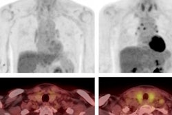
 (A) Single-tracer PET/CT with F-18 FDG showing fused images in the axial plane of a primary tumor at the gastroesophageal junction with a metastasis in the left adrenal gland and liver metastases. (B) Maximum intensity projection (MIP) of single-tracer PET images displaying high uptake in brain tissue, tracer accumulation around the injection side at the right elbow, lymph node metastasis in the left upper mediastinum, multiple abdominal lymph node metastases, and liver metastases. An additional benign accumulation of FDG is visible, caused by right-sided thoracolumbar osteoarthritis. (C) Transverse section of fused dual-tracer F-18 FDG and Ga-68 FAPI-46 PET/CT of the same patient. (D) As all images were visually normalized to the uptake of the liver, MIP of dual-tracer PET/CT in the same patient shows a less pronounced tracer accumulation in the brain tissue, compared to single-tracer PET/CT. In addition to lesions detected with single-tracer PET/CT, further abdominal lymph node metastasis and liver metastases are visualized via dual-tracer PET/CT. The focal tracer accumulation in the right vein angle is due to intravenous tracer accumulation from the former tracer depot at the right elbow. Image courtesy of the Journal of Nuclear Medicine.
Since the diagnostic performance of Ga-68 FAPI-46-PET/CT is best shortly after injection and F-18 FDG-PET/CT is currently the gold standard, the authors so far recommend the injection of Ga-68 FAPI-46 as a second tracer after the F-18 FDG-PET/CT scan.
"Future studies may consider simultaneous injection of both tracers and acquisitions of just one single scan, to further simplify the procedure," Roth and colleagues concluded.