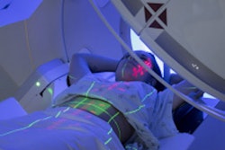
Radiation protection is vital in breast screening exams with x-ray technology, particularly given the increasing use of more complicated and potentially more radiation-intensive procedures such as tomosynthesis and contrast mammography. That's the view of a leading European expert on breast imaging.
 Prof. Dr. Matthias Dietzel.
"This topic is often underestimated or even ignored, and at times, the awareness of our colleagues is alarmingly low. It is simply more exciting to talk about tomosynthesis and contrast-enhanced mammography," Prof. Dr. Matthias Dietzel, from the Department of Radiology at University Hospital Erlangen, Germany, told AuntMinnieEurope.com.
In his role as deputy editor of the European Journal of Radiology, Dietzel is overseeing a virtual special issue called "New Trends in Breast Imaging."
As part of this project, he is planning a short series on radiation protection, patient safety, and the biological effects of ionizing radiation in breast imaging. He is being supported in this task by Prof. Pedro Vaz, PhD, from the Instituto Superior Técnico at the University of Lisbon, Portugal.
Basics of x-ray dosimetry
The first article addresses the issue of x-ray dosimetry in breast cancer screening with 2D and 3D mammography. It was posted online on 28 March and is due to appear in the June 2022 edition.
To estimate the mean glandular dose (MGD), it is important to understand the radiation-associated risk from breast x-ray imaging exams, but this continues to be the subject of much debate because its accuracy is directly related to risk estimation and optimizing breast cancer screening programs, explained first author Salvatore Di Maria, a medical physicist from Centro de Ciências e Tecnologias Nucleares (C2TN) in Lisbon, Portugal, and colleagues.
Recent studies have reported on methods for estimating individual breast glandularity using a software algorithm, permitting a move toward personalized breast dosimetry, but differences in breast density between individual cases could lead to MGD difference of about 13%, especially for thin breasts, according to the authors. The distribution of fibroglandular tissue along the x-ray source to detector direction is unknown, however, and this may result in overestimation of dose.
"The use of 3D breast imaging, together with artificial intelligence, may help to better determine the distribution of glandular tissue and needs investigation. The need for patient-specific dosimetry in mammography could be seen as part of the general patient-dependent dosimetry rationale and a move towards individualized radiological risk assessment," they wrote.
In parallel, scientists are also working toward harmonizing dosimetric protocols and comparing the performance of several mammography systems worldwide. An important initiative is the joint Task Group/Workgroup of the American Association of Physicists in Medicine (AAPM) and the European Federation of Organization for Medical Physics (EFOMP), which aims to improve breast dosimetry models
Basic principles
According to Dietzel, "The authors have done an excellent job of summarizing the basic principles of dosimetry in x-ray examinations in a concise and generally understandable way. Given the subject matter, that's really not easy."
Future articles will focus on specific procedures, including contrast-enhanced mammography, tomosynthesis, and breast CT. All the papers will be available free-of-charge and aim to give a state-of-the-art explanation of all important breast imaging topics.
"Unlike typical special issues, this issue operates on a snowball principle," he said. "There is no deadline, but whenever news seems important to me, I will invite prominent and internationally recognized authors to talk about the topic. This is an ongoing series."
For more details about the virtual special issue, go to the EJR website.