Medical imaging is already playing a valuable clinical role in the Tokyo Olympics, and both MRI and ultrasound look set to be essential diagnostic tools right up to the closing ceremony on 8 August.
Most of the sporting action so far at the Games has been held away from the Olympic Village, which means the bulk of scans have taken place at local hospitals throughout Japan. For instance, when the New Zealand (NZ) captain was injured in the third minute of Sunday's soccer game against Honduras, he was taken to a nearby hospital in Kashima for an MRI exam.
"Our team doctor sent the scan to be read back home," NZ coach Simon Hilton told AuntMinnieEurope.com on 26 July. "The game ended at 6.45 p.m. local time, and by 10 p.m., we knew the outcome was a grade 1 medial collateral ligament injury of the knee."
During a pretournament friendly match at Atsugi, ultrasound was performed on another NZ player. The imaging service has been efficient and speedy, according to Hilton, who noted that keeping the players' professional clubs informed about any injuries is an important task.
Absence of overseas radiologists
There has been some disappointment, however, for some overseas radiologists who were originally due to work at the Games, including Dr. Daichi Hayashi, PhD, assistant professor of clinical radiology at Stony Brook University Hospital, New York.
"Unfortunately, due to the COVID pandemic and associated restrictions in Tokyo, Japan, none of us U.S. radiologists are traveling to Japan for the Olympics," he told AuntMinnieEurope.com.
Hayashi said he was last in touch with Prof. Mamoru Niitsu, professor and chairman of radiology at Saitama Medical University in Japan and chief musculoskeletal (MSK) radiologist for the Tokyo Olympics, in spring 2020. They were working out the logistics of travel to Japan for the Olympics in summer 2020, but the event was postponed for a year due to the worldwide lockdown.
Niitsu is a global expert on knee imaging, and Hayashi was the translator for Niitsu's book, Magnetic Resonance Imaging of the Knee.
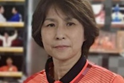
 Prof. Mamoru Niitsu. Photo courtesy of the ESR.
"Precise diagnosis and appropriate treatment are required to enhance the performance of athletes," he told the European Society of Radiology (ESR) in an interview for the 2019 International Day of Radiology, which was devoted to sports imaging.
"As they initiate many of the activities of medical teams, radiologists, who have access to a variety of data, can direct the next steps in treating each patient as well as the future practice of medical teams. Of course, radiologists should provide careful physical and mental support to injured athletes," he added.
Niitsu is a visiting radiologist at the Japan Institute of Sport Sciences (JISS), where training and medical checkups of top athletes, including Olympic athletes, are performed. The Japanese government established JISS in 2001, and the facility has two 3-tesla MRI scanners and other equipment. Delegations from JISS observed at the Olympics held in Athens, Beijing, London, Rio de Janeiro, and Pyeongchang, South Korea.
"Radiologists are often the first point of contact between patients and medical teams," he told the ESR. "All of the activity of medical teams is based on the initial diagnosis. Therefore, the role of radiologists is essential. In addition, radiologists act as coordinators of medical teams, directing further examinations and procedures."
They should monitor patients' ongoing treatment and follow-up and have frequent and close communication with surgeons and other members of staff, he added.
The Japanese system
In Japan, all medical procedures are performed under the national health insurance system or personal health insurance coverage, except on rare occasions, for example when such procedures are performed for JISS or a professional baseball/football team.
"As MRI and CT scanners are in very high demand, all imaging examinations have to be booked in advance. This can result in selection bias for such examinations, but it can also help to exclude unnecessary examinations. Radiologists should recommend the most effective and balanced examination pathway for each patient," Niitsu said.
In the country, marathon running and football have gained in popularity over the past decade, causing a number of injuries and diseases that radiologists must become familiar with. MRI plays a significant role -- and CT, a relatively small role -- in joint and muscle imaging.
"I work at a university hospital with approximately 1,000 beds," Niitsu noted. "I am responsible for reading all MSK images, which account for a fourth to a third of all images read in daily practice. Half of the MSK imaging performed in daily practice is sports-related imaging, for example, in patients with muscle strains, joint injuries, or traumatic bone injuries. The other half involves patients with osteoarthritis, rheumatoid arthritis, myopathy, diabetic foot injuries, etc."
Growing incidence of leg injuries
Contact sports, including rugby, football, and basketball, and events involving long periods of running -- such as marathons, triathlons, and soccer -- are particularly associated with leg injuries, he continued.
With the recent marathon boom, many amateur runners complain of joint pain and muscle soreness. Soccer is the most popular sport among Japanese kids, replacing baseball 10 years ago. As a result, Osgood-Schlatter disease, Sinding-Larsen-Johansson disease, Jumper's knee, and meniscal/ligament injuries now attract much attention.
Recently, focal periphyseal edema and Morel-Lavallee lesions have been included among the differential diagnoses for knee pain in young athletes. In addition, cartilage imaging of the hip, knee, and ankle has become more important, according to Niitsu.
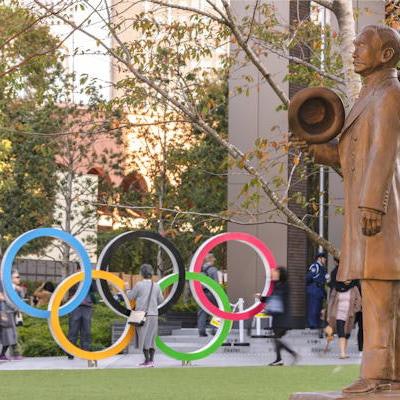
 This statue of the founder of judo, Kano Jigoro, is located in front of the Olympic Rings Monument at the Japan Olympic Museum. Jigoro was the first Asian member of the International Olympic Committee. Photo courtesy of Kuremo / Adobe Stock.
"I mainly diagnose articular cartilage damage. Qualitative MRI techniques can be used to perform internal evaluations, such as assessments of the glycosaminoglycan (GAG) concentration based on T2 mapping, T2* mapping, T1rho mapping (also known as T1ρ or "spin lock;" 'ρ' is the symbol for the Greek letter rho), and chemical exchange saturation transfer (CEST)," he said.
"Cartilage damage can be detected with MRI," he continued. "High-quality MRI images with sufficient spatial and contrast resolution can detect subtle cartilage surface irregularities. Moreover, qualitative imaging, such as T1rho mapping, can be used to evaluate internal changes in the cartilage layer, which makes it possible to assess ongoing changes in the articular cartilage."
Niitsu began working at the MRI Laboratory of the Department of Diagnostic Radiology at the Mayo Clinic in 1991, and he was involved in a number of MR research activities, such as kinematic joint imaging, muscle kinematic MRI and T1 calculation using tag implementation. He continued and expanded MR research, especially in musculoskeletal imaging, after moving back to Tsukuba and Saitama.
Niitsu was president of the Japanese Society for Magnetic Resonance in Medicine (JSMRM) from 2012 to 2014 and chair of the society's annual meeting in 2016 and its international committee from 2016 to 2018.
Editor's note: Prof. Niitsu updated us on 28 July that he is no longer a member of the medical team at the Olympic Village. We hope to post an interview soon with the chief radiologist Dr. Yukihisa Saida.