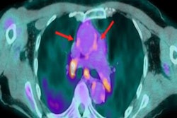
A series of FDG-PET images of COVID-19 patients that shows how the disease causes long-term neurological changes associated with cognitive impairment was given Image of the Year honors at the Society of Nuclear Medicine and Molecular Imaging (SNMMI) meeting.
A group of German researchers from University Medical Center Freiburg in Germany presented images from FDG-PET scans of hospitalized COVID-19 patients with cognitive impairment before and after they recovered. They compared them to images from a normal patient group and found signs of neocortical dysfunction persisted for up to six months.
"As the SARS-CoV-2 pandemic proceeds, it has become increasingly clear that neurocognitive long-term consequences occur not only in severe COVID-19 cases, but in mild and moderate cases as well," said lead study author Ganna Blazhenets, PhD.
Given the rapid spread of SARS-CoV-2 and emerging evidence of neurological symptoms in hospitalized COVID-19 patients, the researchers sought to prospectively assess the disease's neurological effects on FDG-PET scans.
They identified 15 hospitalized COVID-19 patients who showed at least one new symptom of cognitive impairment after taking Montreal Cognitive Assessment tests. After FDG-PET scans, the authors found frontoparietal hypometabolism in 10 of the patients.
Visual interpretation also indicated highly significant (p < 0.001) COVID-19-related spatial covariance patterns characterized by positive weights in brain stem, cerebellum, white matter, and mesiotemporal structures, according to the researchers.
After six months, eight of the COVID-19 patients presented for follow-up FDG-PET scans. In these patients, the mean pattern expression score at the chronic stage was significantly lower compared with the subacute stage (two-tailed paired t-test, p = 0.002), but still higher at trend-level compared with the control group. Only two of the patients had undergone ICU treatment.
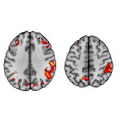
 (A) COVID-19-related spatial covariance pattern of cerebral glucose metabolism overlaid onto an MRI template. Voxels with negative region weights are color coded in cool colors and regions with positive region weights in hot colors. (B) Association between the expression of COVID-19-related covariance pattern and the Montreal Cognitive Assessment (MoCA) score adjusted for years of education. Each dot represents an individual patient. (C) Results of a statistical parametric mapping analysis. Upper row illustrates regions that show significant increases of normalized FDG uptake in COVID-19 patients at six-month follow-up compared with the subacute stage (paired t-test, p < 0.01, false discovery rate-corrected). Bottom row depicts regions that still show significant decreases of normalized FDG uptake in COVID-19 patients at six-month follow-up compared with the age-matched control cohort at an exploratory statistical threshold (two-sample t-test, p < 0.005). Image courtesy of Ganna Blazhenets, et al.
"We can clearly state that a significant recovery of regional neuronal function and cognition occurs for most COVID-19 patients based on the results of this study. However, it is important to recognize the evidence of longer-lasting deficits in neuronal function and accompanying cognitive deficits is still measurable in some patients six months after manifestation of disease," Blazhenets said.
As a result of the study, the authors suggest post-COVID-19 patients with persistent cognitive complaints should be presented to a neurologist and possibly allocated to cognitive rehabilitation programs.
SNMMI's scientific program chair Dr. Umar Mahmood, PhD, who presented the Image of the Year award, said the study provides further evidence that FDG-PET can be applied to unravel neuronal correlates of cognitive decline in COVID-19 patients. Since F-18 FDG-PET is widely available, it may aid in the diagnostic workup and follow-up in patients with persistent cognitive impairment after COVID-19, he stated.
The Image of the Year was chosen from more than 1,280 abstracts submitted to the meeting and voted on by reviewers and the society leadership, the SNMMI said in a statement.