Contrast-enhanced spectral mammography (CESM) is becoming a useful clinical technique due to its relatively low cost, increasing availability, and suitability for women who are contraindicated for MRI, but some important practical drawbacks remain, according to award-winning Spanish research presented at the RSNA 2020 meeting.
CESM provides both morphological and functional information about the study of breast cancer and can achieve better results than conventional digital mammography in the clinical evaluation of dense breasts, noted Dr. Maria Cristina Arízaga Ramirez and colleagues from the breast section of the radiology department at Hospital Clínico San Carlos in Madrid.
"Inconclusive findings, symptomatic patients, and known breast neoplasm are among its main clinical applications," they stated in a digital poster that received a certificate of merit from the RSNA judges "Preliminary studies have shown comparable sensitivity with MRI and a lower false-positive rate when it comes to assessing the extent of breast cancer."
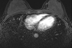
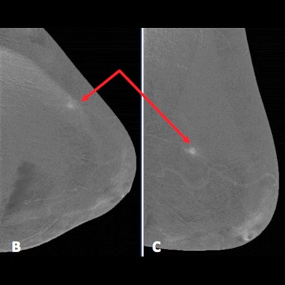
 Above: Mammographic images (A-C) show small enhancing focal asymmetry in upper external quadrant of left breast (red arrows). Below: Ultrasound image shows the enhancement asymmetry corresponds to well-defined hyperechogenic nodule (blue arrows). A biopsy confirmed local inflammatory changes related to fat necrosis. Figure and table courtesy of RSNA 2020 and Dr. M.C. Arízaga, Dr. M. Montés Fernández, et al.
During the authors' first year of using CESM, they scanned 121 patients. The main indication was symptomatic breast disease, and CESM was indicated in a small percentage of patients (4%) due to MRI contraindications.
The most common BI-RADS assignation on diagnostic mammography (low-energy exposure) was 2 (37%). The benign morphological findings (BI-RADS 2), along with the absence of enhancement on high-energy exposure, reinforced the probability of benignity and avoided unnecessary biopsies. Of the BI-RADS 4 and 5 cases (44%), malignancy was confirmed in 78% of patients, they reported, adding that a comprehensive and multidisciplinary diagnostic approach is essential.
Problem-solving
Women at high risk of developing cancer may go undetected in conventional mammography breast screening due to its low sensitivity, explained Arízaga Ramirez, who is a third-year resident. Dr. Myriam Montés Fernández, the radiologist in charge of the breast section in Madrid, was a co-author of the RSNA poster.
Those with genetic risk (BRCA1 and BRCA2 mutations) undergo MRI as a screening technique, but there are other risk factors that do not qualify a patient for an MRI screening, such as family and personal history of breast cancer and dense breast pattern, the researchers noted.
"In dense breasts, the specificity of digital mammography is low, therefore many asymmetries, distortions, or even masses can go unnoticed," they pointed out. "Contrast mammography will solve this problem by highlighting pathological areas of contrast uptake not seen on diagnostic mammography."
Specificity and sensitivity increased by 4% and 45% respectively within experienced radiologists and by 3% and 30% respectively with experienced and inexperienced readers.
Benefits and drawbacks
To determine the existence of other associated lesions, additional techniques such as ultrasound and MRI may be requested when a primary breast tumor is diagnosed. MRI is the standard reference to assess the extent of the disease. Research shows that CESM is sensitive in the detection of other multicentric, multifocal, or bilateral lesions, although to a lesser degree than MRI for ipsilateral lesions, and false negatives have been found when the lesion is not included in the field-of-view, the authors wrote.
Compared with MRI, CESM has fewer false positives, and therefore better specificity, but its use has not been standardized due to the lack of experience in many centers and because it is still undergoing study. In patients who cannot clearly undergo MRI, CESM is an adequate option, they noted.
Breast cancer is the most common malignant pathology in women, and 1 out of 8 women are known to have an individual lifetime risk of developing breast cancer. MRI is still the gold standard test for providing information related to neovascularization, such as a specific tumor characteristic, improving the detection and characterization of breast cancers, but CESM is showing promise and can contribute to the characterization of malignant lesions with a sensitivity rate comparable to MRI, according to the Madrid team.
Malignant lesions tend to show intense enhancement -- due to their hypervascularization -- and also tend to present spiculated margins. CESM helps to detect other associated lesions in the same or contralateral breast.
Benign lesions are highly prevalent and in many cases, they share radiological similarities with malignant processes, and it is important for the radiologist to know how to distinguish them and to correlate the radiological appearance with a patient's risk factors, the researchers continued.
Many malignant entities present enhancement in CESM as they are hypervascular, but there are benign diseases that can also enhance and therefore present as false positives. A tool that can reduce false positives is the kinetic assessment of contrast uptake curves, which in CESM are not entirely comparable with those of MRI, they wrote.
Neoadjuvant systemic therapy
In recent years, neoadjuvant systemic treatment has become much more important in locally advanced cancers. And for surgical planning, it is essential to have an adequate imaging assessment of the response to treatment. MRI is the technique of choice for this, given its acceptable sensitivity and specificity, but in some studies, CESM has shown sensitivity, specificity, and positive and negative predictive values only slightly lower than MRI, so it could become an alternative in future years after more investigations, the authors stated.
CESM had 100% sensitivity in detecting a complete pathological response, but a lower specificity at 67%, with six false negatives lowering the overall accuracy of the technique.
MRI has several techniques such as diffusion or contrast kinetic curves that allow detection of findings before there is a visible reduction in size -- something that is not possible even with CESM.