Abdominal sessions at ECR 2020 in July are set to feature many highlights, including new advances in pancreatic imaging, the use of imaging to predict therapeutic success, and how best to optimize and shorten imaging protocols, according to Prof. Dr. Thomas Lauenstein from Düsseldorf, Germany.
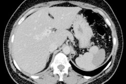
In pancreatic imaging, the latest research will show why imaging surveillance is suitable for slow-growing cystic pancreatic lesions, he noted in an editorial published online 20 May in European Radiology.
 Dr. Thomas Lauenstein.
"However, radiologists should be aware of risk factors that may lead to a change of the surveillance rate," he pointed out. "These factors include patients' age, number of lesions, presence of septa, and visible communication with the pancreatic duct."
Other abdominal imaging presentations will explore the potential of multiparametric imaging for predicting treatment outcomes in tumor patients.
"Imaging features may help to answer the question whether a specific treatment may be successful or if other alternatives should be taken into consideration," he stated. "Furthermore, it may be a guideline to tailor surveillance strategies: If pretherapeutic imaging reveals high risk of tumor recurrence, time intervals for follow-up imaging need to be adapted."
However, challenging issues still remain, including questions over the practicability of these prediction strategies and the use of different imaging modalities and imaging parameters in these studies, according to Lauenstein.
"Hence, it would be advantageous to synchronize results in order to allow for a more uniform implementation," he added.
Other abdominal imaging studies will describe advances for optimizing and shortening imaging protocols. Lauenstein said that the evaluation of short MRI protocols is promising and will encourage radiologists to tailor examinations for specific clinical circumstances.
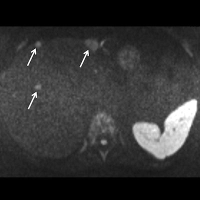
 Obtained during a short protocol, this diffusion-weighted image shows a patient with conspicuous liver metastasis. The lesions are marked by arrows. Courtesy of Dr. Thomas Lauenstein. Originally published in the ECR Today newspaper.
"Hence, a dedicated first-time MRI exam of the liver could encompass more or different sequences than a follow-up exam or a surveillance exam of patients with a high-risk profile," he explained. "If we follow this strategy, examination time can be spared and more exams per time unit could be performed."
Some problems still need to be resolved, however, including when to use intravenous contrast and the somewhat heterogeneous nature of the proposed short MRI protocols.
"It will be the task of expert groups or multicenter trials to overcome this limitation in the future," he wrote.