Swiss researchers have identified several key MRI-based indicators that can help determine how well pediatric patients will recover from a brain injury. They published their study findings online on 19 March in the European Journal of Radiology.
The degree of brain injury and loss of white-matter volume combined with fractional anisotropy values within the corpus callosum could be used to best predict how well youngsters would respond to rehabilitation. Patients in the study tended to have better outcomes if they were older and did not spend as much time in rehabilitation.
"These results support the utility of MRI measures as predictors for cognitive outcome in patients with acquired brain injury, and highlight in particular the importance of the corpus callosum for cognitive function, thus adding to the growing body of knowledge, which describes the role of the corpus callosum in cognition," explained first author Volker Ressel, PhD, from University Children's Hospital Zurich and colleagues.
MRI has been the primary modality for assessing traumatic brain injuries and strokes due to its high sensitivity, compared with other modalities. Diffusion-tensor MRI (DTI-MRI) has proven particularly useful with its ability to evaluate the brain's white matter and the corpus callosum, which directs communication between the right and left hemispheres.
"In children, abnormal callosal development can lead to deficits in functional connectivity that are related to impairments in specific cognitive domains, showing that the corpus callosum plays an important role in the development of cognition," the authors noted. Therefore, it is important to accurately evaluate this region of the brain after an injury to chart the best rehabilitative plan for young patients.
For this retrospective study, Ressel and colleagues analyzed data from 21 children (mean age, 11.6 years; range 7.1-19.4 years) who had either a stroke or traumatic brain injury and had undergone a structural 3-tesla MRI scan with DTI (Signa HDxt or MR750, GE Healthcare) prior to rehabilitation.
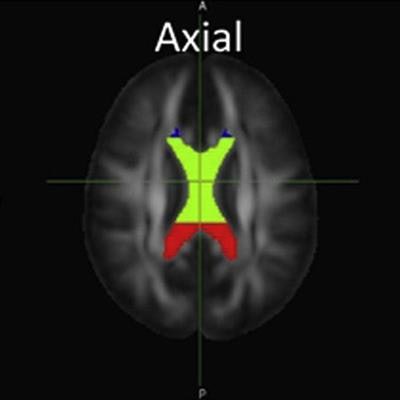
 DTI-MR images are examples of the corpus callosum and its substructures -- genu (blue), body (green), and splenium (red) -- used in this study, as defined by software from Johns Hopkins University. Image courtesy of the European Journal of Radiology.
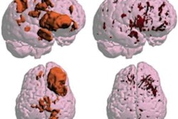
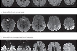
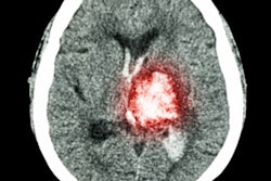
Through DTI, the researchers targeted mean fractional anisotropy values in the corpus callosum and its substructures -- genu, body, and splenium -- which are believed to serve an important role in cognition and neural communication. Structural MR images were used to score the severity of brain injuries based on the number of lobes affected, volume and type of white-matter injury, and grey-matter damage.
To quantify the efficacy of rehabilitation, the researchers used a pediatric version of the Functional Independence Measure (WeeFIM) scoring model, focusing specifically on comprehension, expression, social interaction, problem solving, and memory. They considered a WeeFIM cognitive score greater than 30 to be a good outcome. The team calculated sensitivity, specificity, and area under the curve (AUC) of the mean fractional anisotropy and MRI injury scores using a receiver operating characteristic (ROC) analysis.
Comparisons of all the variables determined that the combination of mean fractional anisotropy within the corpus callosum body and the brain injury score provided the highest sensitivity (86%), whereas the mean fractional anisotropy within the corpus callosum splenium showed the highest specificity (100%). The patient's age at the time of rehabilitation and amount of time spent in the program also influenced cognitive recovery significantly.
"Specifically, patients tend to have better cognitive outcomes if they are older and spend less time in rehabilitation, but the latter factor is likely to reflect a lower severity of the underlying injury, or a result of a better post-injury recovery," Ressel and colleagues wrote.