Swiss researchers have used cinematic rendering to create photorealistic images of head CT and conebeam CT scans. They assess the potential benefits of integrating these images into the evaluation of maxillofacial anatomy and pathology in a study recently published online in Dentomaxillofacial Radiology.
Clinicians are increasingly turning to 3D image reconstruction to facilitate diagnosis and treatment planning for a variety of medical conditions. In the case of maxillofacial lesions, use of conventional volume rendering is common and also widely considered beneficial for detection and interpretation, according to study co-authors Drs. Sebastian Winklhofer and Bernd Stadlinger, and colleagues from the University of Zurich.
More recently, several groups have explored using an advanced reconstruction technique known as cinematic rendering to further enhance the visualization of complex anatomy, primarily for highly vascularized structures such as the pancreas, colon, and spleen. (Cinematic rendering involves applying a global lighting model to medical images to generate photorealistic 3D reconstructions.)
"Imaging techniques like cinematic rendering can help to increase the ability for an improved 3D understanding and imagination," the authors wrote. "Introducing such techniques to radiology facilitates the approach to read radiological images."
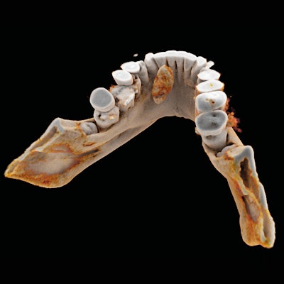
In the current study, Winklhofer, Stadlinger, and colleagues used computer software (syngo.via Frontier VB30, Siemens Healthineers) to create cinematically rendered images based on patients' CT and conebeam CT scans. The technique enabled them to visualize high-density structures such as bones, teeth, and tissue calcifications in great detail (Dentomaxillofac Radiol, 8 August 2019).
The team of radiologists and surgeons examined various types of health complications affecting the face and the mouth of several patients, including the following:
- Jaw malformation: Cinematically rendered images allowed the group to visualize at precisely what point the upper and lower teeth of a patient with dysgnathia complex were unable to make contact upon closing.
- Jaw fracture: Conventional volume rendering offered a limited view of a lateral jaw fracture that was much more readily noticeable with cinematic rendering due to the technique's added spatial depth.
- Post-traumatic tooth fracture: Cinematic rendering showed the surface of the tooth and fracture, as well as the extent to which the fracture line invaded the tooth's center, or pulp cavity.
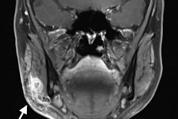
- Salivary gland stones: Though a patient's salivary gland stones were identifiable on standard conebeam CT, cinematically rendered images revealed that the stones were much closer to the jaw than previously believed -- highlighting the technique's possible utility in presurgical planning.
 Patient with a salivary gland stone visualized with axial conebeam CT (a), conventional volume-rendering reconstruction of conebeam CT scans (b, c), and cinematic rendering of conebeam CT scans (d-f). Image courtesy of Drs. Sebastian Winklhofer and Bernd Stadlinger.
"Cinematic rendering is a helpful tool for a better understanding of complex spatial anatomical structures and enables a facilitated demonstration of pathologies not only to medical doctors but also to nonmedical professionals, in particular, to patients," the authors wrote.
The highly detailed, photorealistic appearance of cinematic rendering reconstructions makes it ideal for teaching and training students and residents as well, they noted.