Patients receiving stereotactic body radiotherapy (SBRT) for central lung tumors face an increased risk of central airway toxicity, which in some cases may be potentially life threatening. Markerless 3D monitoring of the proximal bronchial tree could help manage this risk by tracking the position and motion of airways relative to the tumor, while offering a more reliable tracking option than reflective or implanted markers. Accurate, real-time position monitoring could alert users to interrupt radiation treatment if the proximal bronchial tree position deviates beyond a predetermined threshold.
Researchers at the VU University Medical Center (VUmc) in Amsterdam have investigated proximal bronchial tree motion monitoring using a markerless template matching and triangulation technique, based on kV projection images acquired during linac gantry rotation. Their study demonstrated the technique is technically feasible for use during central lung SBRT (Radiotherapy and Oncology, 29 August 2018).
Lead investigators Max Dahele and Wilko Verbakel from VUmc's department of radiation oncology, together with doctoral student Colien Hazelaar and colleagues, conducted a feasibility study of markerless 3D position monitoring using kV projection images, which offer the capability for simultaneous visualization of tumor and airway position. They examined conebeam CT (CBCT) projections of a 3D-printed thorax phantom and clinical data, and employed prototype software for template generation, template matching, and triangulation to determine bronchus motion in three dimensions.
Four-dimensional CT is used for treatment planning to determine a patient's breathing motion and relative positions of organs-at-risk (OAR). However, these positions may change during the actual treatment. Internal and external markers can be used to monitor motion during treatment, but any marker is just a surrogate and may not capture the actual motion of the airway. This is not the case with real-time markerless position monitoring of the airways themselves.
Matching motion
The researchers created a 3D-printed phantom consisting of soft tissue, bony structures, airways, lungs with blood vessels, and three lung tumors. They simulated irregular breathing motion by automatically moving the treatment couch, and acquired kV images for a full arc at 15 frames/second. Two structures, representing the left and right proximal bronchial trees, were delineated on the planning CT scan. They also performed fluoroscopy during MV irradiation, acquiring images at 7 frames/second.
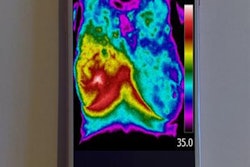
 A typical trace shows the motion of a patient's airway. Image courtesy of Wilko Verbakel.
In addition, the researchers retrospectively analysed full-fan CBCT images (without irradiation, with 470-500 images per dataset) from 10 patients who had undergone free-breathing stereotactic or hypofractionated lung irradiation. All patients had tumors located within the field-of-view of a full-fan CBCT scan, with half of the tumors on the right side of the lung. The proximal bronchial trees were similarly delineated.
The authors used prototype software (template-based tracking and the Sequential Stereo algorithm) to create 2D reference templates for every degree of gantry rotation. The templates included the delineated airways plus a 4 mm isotropic margin to include the airway walls. They selected the template associated with the gantry angle closest to the projection image. They matched the templates to the projection images for 2D proximal bronchial tree position using normalized cross-correlation; and used multiple registrations triangulated to determine the 3D position.
The right proximal bronchial tree had a better matching performance than the left proximal bronchial tree, which the authors attributed to the mediastinum being more obscured on the left side. In the phantom, the 2D right/left proximal bronchial tree position could be determined in 86.6%/75.1% of the CBCT datasets without MV irradiation; the 3D position (excluding the first 20°) was determined in 84.7%/72.7% of datasets.
In the patient dataset, there were no remarkable differences in matching performance between the left and right proximal bronchial tree, with 2D position determined in 89.8% of each dataset, 3D position in 76.4%, and 3D position (excluding the first 20°) in 85.1%. The authors suggested that performance could potentially be improved by analyzing more data and creating more optimal triangulation parameter settings. They note that dynamic adjustment of kV and mA might offset poorer results of template matching when image quality was poor. They also advised that a different structure with distinguishable features could be used, such as branches of the proximal bronchial tree.
"We are currently using the same software for continuous spine position tracking during spine SBRT delivery," Verbakel said. "It is updated subsecond, and the accuracy is in the order of 0.3 mm. If the spine position during radiation delivery is more than 1 mm off, we interrupt the treatment for a new setup CBCT scan."
"We can also use the software for lung tumor position monitoring during breath-hold lung SBRT delivery," he explained. "Because there can be large inter-breath-hold variation, we like to monitor if the tumor is indeed within the PTV. In case the tumor moves outside the PTV, we interrupt the treatment and do a new breath-hold, or a new setup CBCT scan."
© IOP Publishing Limited. Republished with permission from Physics World, a website that helps scientists working in academic and industrial research stay up to date with the latest breakthroughs in physics and interdisciplinary science.